Clin Mol Hepatol > Volume 28(1); 2022 > Article
Lee and Seo: Current knowledge about biomarkers of acute kidney injury in liver cirrhosis

ABSTRACT

Acute kidney injury (AKI) is common in advanced cirrhosis. Prerenal azotemia, hepatorenal syndrome, and acute tubular necrosis are the main causes of AKI in patients with cirrhosis. Evaluation of renal function and differentiation between functional and structural kidney injury are important issues in the management of cirrhosis. However, AKI in cirrhosis exists as a complex clinical spectrum rather than concrete clinical entity. Based on current evidence, changes in serum creatinine (Cr) levels remain the most appropriate standard for defining AKI in cirrhosis. However, serum Cr has a limited role in assessing renal function in this population. This review examines previous studies that investigated the ability of recent biomarkers for AKI in cirrhosis from the perspective of earlier and accurate diagnosis, classification of AKI phenotype, and prediction of clinical outcomes. Serum cystatin C and urine neutrophil gelatinase-associated lipocalin have been extensively studied in cirrhosis, and have facilitated improved diagnosis and prognosis prediction in patients with AKI. In addition, urine N-acetyl-β-D-glucosaminidase, interleukin 18, and kidney injury molecule 1 are other promising biomarkers for advanced cirrhosis. However, the clinical significance of these markers remains unclear because there are no cut-off values defining the normal range and differentiating phenotypes of AKI. In addition, AKI has been defined in terms of serum Cr, and renal biopsy—the gold standard—has not been carried out in most studies. Further discovery of innovate biomarkers and incorporation of various markers could improve the diagnosis and prognosis prediction of AKI, and will translate into meaningful improvements in patient outcomes.

INTRODUCTION

Renal dysfunction is a frequent and life-threatening complication of advanced cirrhosis [1-3]. It has been established that acute kidney injury (AKI) directly correlates with the prognosis of patients with cirrhosis [4-7]. Furthermore, a gradual increase in serum creatinine (Cr) was observed after the resolution of AKI, and the survival of patients with AKI was significantly reduced compared to that of patients without AKI [8]. AKI in cirrhosis is not a single disease entity; rather, it consists of complex pathophysiology. Hepatorenal syndrome (HRS) is a characteristic feature of advanced cirrhosis; however, it is not the only cause of AKI in these patients [9,10]. Accurate estimation of renal function is important for decision making including liver transplantation (LT). In addition, clearly differentiating the phenotype of AKI in cirrhosis is crucial, because the management of AKI is dictated by its cause.
One of the major limitations in managing patients with AKI and cirrhosis is insufficient ability of serum Cr to act as a kidney biomarker. Serum Cr usually overestimates the renal function in cirrhotic patients who have the high prevalence of sarcopenia [11]. In addition, because Cr is a marker reflecting kidney filtration, not injury, it is not suitable for differentiating the AKI phenotype. Overall, there is a distinct need for reliable biomarkers that can precisely assess renal function and differentiate the phenotypes of AKI in cirrhosis. Novel kidney biomarkers have been evaluated for AKI and cirrhosis to improve both diagnosis and clinical outcomes of the affected patients. This review examines the role of the current biomarkers in assessing renal function, predicting prognosis, and identifying the cause of kidney dysfunction.

DEFINITION AND PHENOTYPES OF AKI

AKI is a spectrum of clinical syndrome, including various precipitating factors that cause either direct injury to the kidney (structural injury) or cause acute dysfunction (functional injury). Risk, injury, failure, loss, end-stage kidney disease (RIFLE), and acute kidney injury network (AKIN) classifications based on changes in Cr and urine output were useful in predicting the prognosis of patients with liver cirrhosis [6,12-17]. In 2012, Kidney Disease: Improving Global Outcomes (KDIGO) proposed the new AKI definition; 1) increase in serum Cr by ≥0.3 mg/dL within 48 hours; or 2) increase in serum Cr to ≥1.5-fold of baseline, which is known or presumed to have occurred within the prior 7 days; or 3) urine volume <0.5 mL/kg/hour for 6 hours [18]. Several studies have validated this definition in hospitalized patients with cirrhosis [6,17,19,20]. However, in cirrhosis, urine volume could decrease without renal dysfunction or increase with the administration of diuretics [21]. Therefore, in 2015 International Club of Ascites consensus, AKI was defined using KDIGO serum Cr criteria, but urine volume was excluded from definition. A study with critically ill patients with cirrhosis showed that KDIGO criteria had higher predictability than RIFLE or AKIN criteria for assessing prognosis in these patients [22].
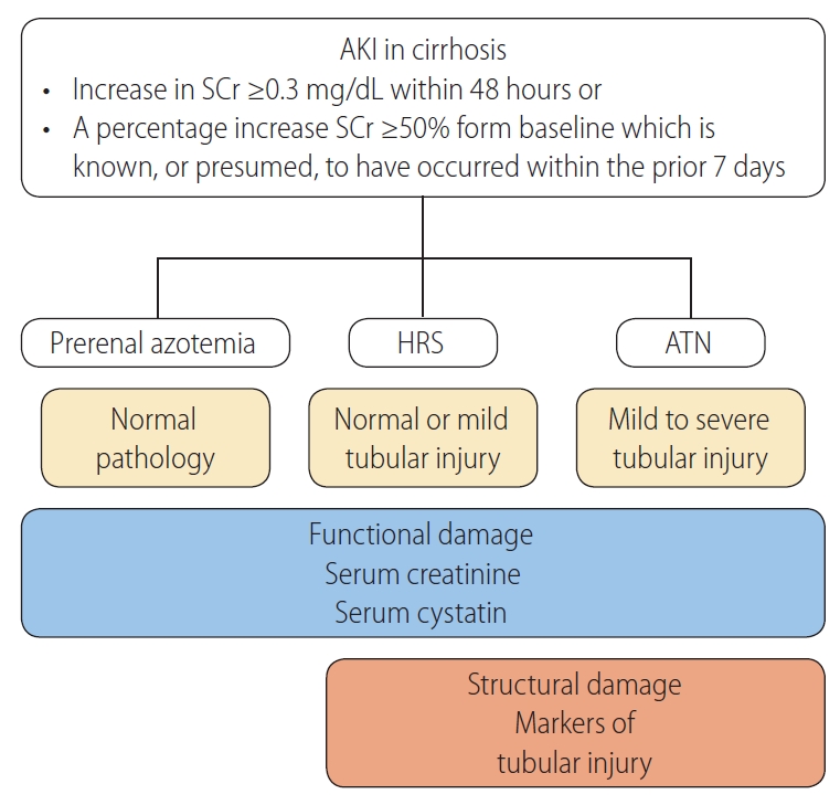
AKI phenotypes are classified as prerenal, intrarenal, and postrenal in the general population. Patients with cirrhosis characteristically develop a specific type of renal dysfunction, HRS [23]. Prerenal types represent approximately two-thirds of cases, whereas intrarenal AKI accounts for one-third of AKI cases, and postrenal AKI is rare in patients with cirrhosis [24]. Reversible AKI in cirrhosis is predominantly a prerenal injury, which resolves with volume administration and discontinuation of diuretics. In contrast, HRS is characterized by its nonresponsiveness to volume expansion [4]. HRS typically represents a continuum of disease, starting with functional changes, followed by structural changes due to prolonged ischemic injury. Intrinsic AKI involves acute tubular necrosis (ATN), acute interstitial nephritis, acute glomerular and vasculitic renal diseases. ATN is a condition that causes a lack of oxygen and blood flow to the kidneys, leading to damaged tubules. Proximal tubule is located in a vulnerable area exposed to hypoxic injury, and patchy areas of flattened tubular cells with enlarged lumens and apical vacuolization are the characteristic features of ATN. Renal tubules can also be injured with ischemia induced by prolonged hypoperfusion. Diagnostic approach of AKI in cirrhosis are presented in Figure 1. Accurately assessing renal function and differentiating the phenotypes of AKI are crucial in managing patients with cirrhosis. Although there have been many efforts, still there are many challenges and needs for relevant renal biomarkers for patients with cirrhosis.

PATHOPHYSIOLOGY OF HRS

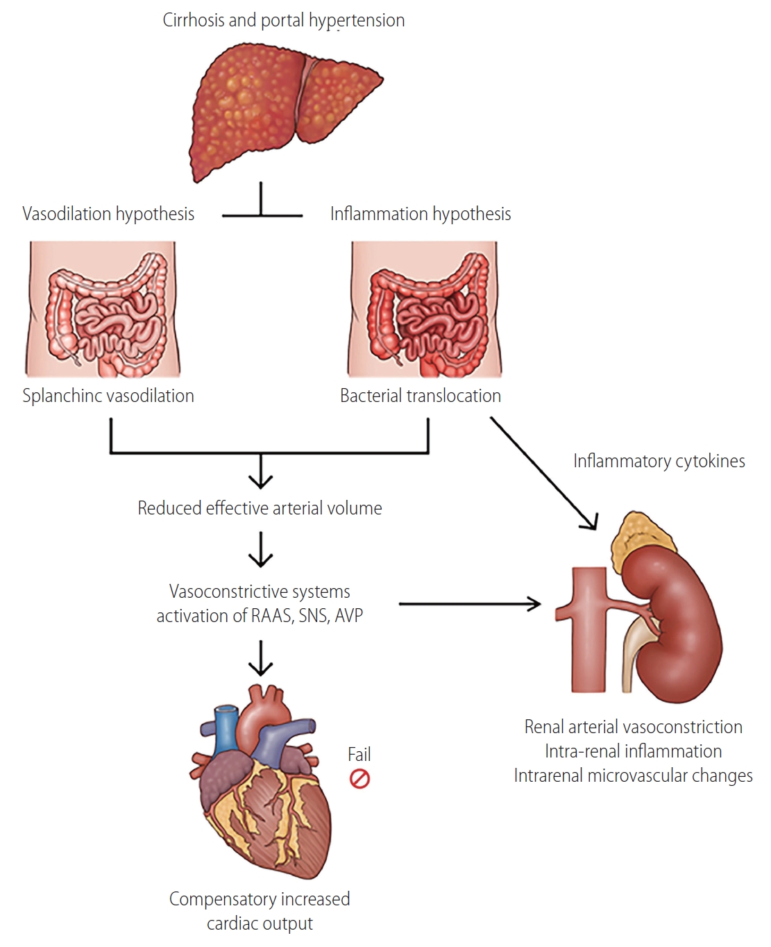
The pathophysiology of HRS comprises several mechanisms, including hemodynamic alteration, kidney factors, and systemic inflammation (Fig. 2) [9,23]. Hypoperfusion plays a major role in the development of HRS. In early-stage cirrhosis, splanchnic vasodilatation results in a decrease in effective arterial volume, and it is balanced by hyperdynamic cardiac compensation to maintain adequate renal perfusion. In more advanced cirrhosis, the renin-angiotensin-aldosterone system, sympathetic nervous system, and arginine vasopressin are activated, leading to systemic vasoconstriction and sodium and water retention in the kidney [2,8]. At most of these stages, increased cardiac output fails to compensate, and this may contribute to a remarkable decrease in renal blood flow [25]. Impaired end-organ perfusion promotes functional kidney injury and a decrease in glomerular filtration rate (GFR), although there is no or minimal parenchymal kidney damage [26]. In addition, in response to changes in the systemic circulation, autoregulation of the renal blood flow by protective mediators could be impaired, further decreasing GFR [27].
Recently, it has been recognized that HRS also involves systemic inflammation [28]. Gut permeability is easily disrupted in patients with cirrhosis, resulting in bacterial translocation. This induces the upregulation of systemic inflammatory mediators, leading to compromised extrahepatic organ perfusion, including the kidney [28-32]. In patients with bacterial translocation, pathogen-associated molecular patterns, such as endotoxins and bacterial DNA, activates monocytes, which leads to the release of pro-inflammatory cytokines such as tumor necrosis factor-alpha, interleukin (IL) 6, and IL-1 beta [33,34]. In addition, increased vasoactive mediators, such as nitric oxide, were observed in these patients. In previous studies, these cytokines were associated with impaired renal function in patients with cirrhosis, as well as in those with acute-on-chronic liver failure (ACLF) and acute liver failure [35,36].
It has been shown in both experimental and clinical studies that the renal tubular toll-like receptor 4 (TLR-4) is overexpressed following inflammatory insult in both experimental and clinical studies [37,38]. Upregulation of TLR-4 is related to the development of tubular cell damage, suggesting that structural changes in the kidney could also contribute to renal dysfunction in HRS. In an experimental study of rats with cirrhosis, tubular injury was observed after the administration of a sublethal dose of lipopolysaccharide, indicating that TLR-4 overexpression is likely a result of bacterial translocation [37]. In patients with sepsis-associated AKI, systemic inflammation-induced tubular cell injury is characterized by vacuolar degeneration, loss of polarity, loss of tight junctions, apoptosis, dedifferentiation and cell cycle arrest [39,40]. According to the results of recent studies, it has been suggested that structural damage could be combined in HRS, which is known to have predominant functional damage.

KIDNEY BIOMARKERS

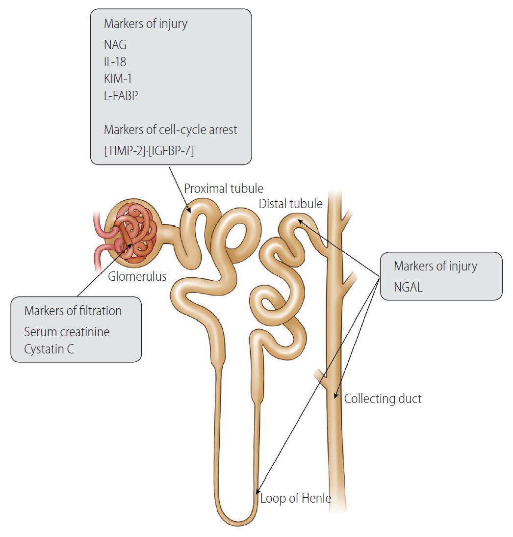
With advances in proteomic platforms, several novel biomarkers of AKI have been discovered [41]. These biomarkers are used in preclinical models of drug development, for early recognition of nephrotoxicity [42]. In clinical studies, kidney biomarkers have been mainly assessed in intensive care unit patients and cardiac surgery populations, who have a high prevalence of AKI [43]. The ability of innovate kidney biomarkers has been investigated from several perspectives. Large prospective studies have demonstrated multiple biomarkers that can detect AKI earlier than could the previous markers [44,45]. Timely identification of AKI using these markers allows for the rapid management of and improved clinical outcomes in the affected populations. Several biomarkers are associated with the progression to a more severe stage of AKI, or long-term prognosis including the development of AKI, reversibility of renal impairment, and mortality [46-50]. Lastly, some biomarkers were useful in differentiating prerenal AKI from ATN, reducing unnecessary volume administration to those with fluid-unresponsive AKI [51,52]. Kidney markers in cirrhosis can be categorized into three groups: 1) markers of kidney dysfunction; 2) tubular injury markers increase with cellular damage, characterized by alteration in cell metabolism and expression of adhesion molecules; and 3) cell-cycle arrest marker detects cellular stress, which is potentially reversible and may or may not develop cell damage (Table 1) [53]. The summary of studies regarding novel kidney biomarkers are presented in Figure 3 and Table 2.

BIOMARKERS FOR THE ASSESSMENT OF KIDNEY FUNCTION

Cr

Cr and Cr-based equations are the most widely used tools for estimating renal function in patients with liver cirrhosis. However, it is well established that serum Cr is not an accurate marker of renal dysfunction in cirrhosis [54-58]. Serum Cr levels vary with sex and age [57]. In some patients, the concentrations of serum Cr remain within normal limits, even in moderate to severe renal dysfunction, resulting in an overestimation of GFR [58-62]. Cr is produced in the liver and stored in the muscle, thus, its serum concentration is intimately related to liver disease and the amount of muscle mass [56,58]. In patients with hepatic dysfunction, the production of creatine, the precursor of serum Cr, is decreased, and tubular secretion of Cr is increased. Sarcopenia, which is prevalent in patients with advanced cirrhosis, also contributes to low serum Cr levels. Therefore, serum Cr level is insufficient for the detection of early-stage renal dysfunction in patients with cirrhosis, which could delay the timely management of kidney injury [60,63].
Inevitably, serum Cr level-derived equations (Cockcroft, modification of diet in renal disease [MDRD]-4, and Chronic Kidney Disease Epidemiology Collaboration equations) tend to overestimate GFR in patients with cirrhosis [11,64-66]. In addition, most serum Cr level-derived equations were developed in patients without cirrhosis, limiting its accuracy in cirrhosis [67-71]. In a large cohort study, MDRD-estimated GFR was overestimated in 47% of patients with liver cirrhosis, compared to the measured GFR, and female sex, advanced cirrhosis, and decreased skeletal muscle mass were independent predictors of overestimation [11]. Other studies have reported that Cr-based equations overestimate renal function by 30–50%, especially in patients with impaired liver function and low GFR [65-67,72].
Conflicting data have been reported regarding the association between Cr levels and the prognosis of cirrhosis. In some studies, a statistical significance between Cr and survival was observed, however, serum Cr levels were not significantly associated with mortality and or the development of HRS in other studies [60,72-75]. In addition, the observed mortality rate of patients with HRS was higher than that predicted by model for end-stage liver disease (MELD) or MELD sodium scores, which estimate renal function by serum Cr levels [76]. Currently, GFR is estimated using the Cr-based MDRD-6 equation in simultaneous liver-kidney transplant guidelines. Unfortunately, the measured GFR was underestimated when the measured GFR was greater than 30 mL/min/1.73 m2 in patients with cirrhosis, which could result in unnecessary simultaneous liver-kidney transplantation (SLKT) [77]. In contrast, the overestimation of the measured GFR could lead to increased mortality after LT [77,78].

Cystatin C

Serum cystatin C has been investigated to improve the detection of reduced GFR [79]. Cystatin C is a 13-kD endogenous cysteine proteinase inhibitor produced at a constant rate by all nucleated cells [80]. Cystatin C is eliminated mainly by glomerular filtration, and is almost completely reabsorbed and catabolized in tubules [81]. Serum cystatin C is not affected by sex, hepatic function, or muscle mass [55,82-85]. The kinetics of serum cystatin C are similar to those of Cr, with the serum concentration increasing 12–24 hours after the onset of AKI [86].
The accuracy of cystatin C-based estimated GFR was compared with that of Cr-based estimated GFR. In a study of 779 patients with cirrhosis, cystatin C-based estimated GFR showed a better correlation with measured GFR than the MDRD-estimated GFR [11]. Other studies demonstrated that Cr-cystatin C combined GFR equations were more accurate than Cr-based equations in predicting measured GFR in patients with cirrhosis, especially when the GFR was lower than 60 mL/min/1.73 m2 in both, the patients with and without ascites [87,88]. In patients with normal range of serum Cr levels, cystatin C was the only independent predictor for the measured GFR [55].
Cystatin C was more accurate than Cr for predicting outcomes of cirrhosis. In patients with cirrhosis, baseline cystatin C-based GFR (cut-off <55 mL/min/1.73 m2) was useful in the early recognition of AKI. In a prospective study of patients with cirrhosis, cystatin C was better at predicting overall survival and the incidence of AKI than MDRD-estimated GFR [11]. Other prospective study of 531 patients with cirrhosis, cystatin C level but not serum Cr was an independent predictor of AKI development, an MELD-cystatin score accurately predicted the AKI development and mortality [89]. In patients with cirrhotic ascites, cystatin C was an independent predictor of mortality and the development of HRS, while Cr was not [75]. Several studies have suggested that changes in cystatin C levels are associated with the development of AKI, however, these are insufficient for supporting the role of change in cystatin C level in defining AKI in cirrhosis [90,91].

BIOMARKERS FOR DEFINING THE PHENOTYPE OF AKI

Identification of AKI phenotype is crucial for establishing a treatment strategy and improving clinical outcomes. Because a clear objective test for HRS is absent, 48 hours of empirical volume expansion is required for both a diagnostic and therapeutic challenges. AKI caused by HRS and ATN persists after volume administration and requires appropriate management for each entity. However, differentiating HRS from ATN is challenging. The major treatment for HRS is the administration of vasoconstrictors, which has been shown to improves the renal function in approximately 50% of the patients with HRS [92-94]. However, short-term mortality of HRS remains very high, up to 90%, and LT is the only treatment that improves the long-term survival of patients with HRS [17,92,95]. In contrast, treatment of the underlying cause is the main therapeutic strategy for ATN. A fluid-first treatment may delay the initiation of vasoconstrictors in patients with HRS, or induce volume overload in those with ATN, who are unlikely to respond, resulting in a poor clinical outcome [96,97].
The level or change in serum Cr cannot differentiate between functional and structural renal impairment [98]. Fractional excretion of filtered sodium (FENa) is used to differentiate the phenotypes of AKI, characterized by FENa less than 1% in prerenal azotemia. However, low FENa can be observed in AKI subtypes other than the prerenal type because of activated water and sodium retention [23,99]. In addition, FENa can be increased by diuretics, glycosuria or chronic adaptation [100]. Proteinuria of over 1–2 g/day suggests a glomerular injury; however, in patients with cirrhosis, serum protein concentration is decreased by impaired hepatic function and poor nutrition, leading to a lower amount of proteinuria [18]. Glomerular injury can be suspected in patients with proteinuria of less than 0.5 g/day when their serum protein level is low. In addition, a poor correlation between conventional biomarkers and kidney histology was observed in a previous study [101]. Because of the disappointing performance of the previous markers in assisting with the differentiation of HRS from ATN and other kidney diseases, there is a great need to identify markers specific to parenchymal kidney insult. Differentiating functional changes from structural changes could help differentiate between patients with a reversible disease and those with irreversible changes.

Tubular injury biomarkers

Tubular injury biomarkers have been shown to have the potential to differentiate HRS from ATN in patients with cirrhosis. Hypoxia leads to proximal tubule dysfunction, resulting in increased excretion of low-molecular-weight proteins into urine or serum. Since they are expressed only hours after kidney injury, they have suggested as the earliest markers of AKI [102-104]. However, there are insufficient data to support their role as early detection markers of AKI in cirrhosis.
The levels of urinary tubular markers are different between AKI phenotypes, with the lowest levels observed in prerenal AKI, modest levels exhibited in HRS, and significantly higher levels found in ATN [105-110]. The potential tubular injury biomarkers in cirrhosis are 1) neutrophil gelatinase-associated lipocalin (NGAL), 2) N-acetyl-β-D-glucosaminidase (NAG), 3) IL-18, 4) kidney injury molecule 1 (KIM-1), and 5) liver-type fatty acid-binding protein (L-FABP).
NGAL is a low-molecular-weight protein (25 kDa) produced by thick ascending and collecting ductal cells of kidney and cells of liver, lung, and gastrointestinal tract [111]. In animal models, NGAL is highly expressed in the kidney and released into the urine after ischemic or nephrotoxic insults. Following ischemic insult, urine NGAL concentration increases within 2 hours [111]. In addition, NGAL plays a role in the innate immune system through iron sequestration [112,113]. Although both serum and urine NGAL can be measured, the serum NGAL levels have not been widely studied.
NAG originates from the lysosome of proximal tubules; therefore, increased urinary NAG levels represent proximal tubule injury with loss of lysosomal integrity [114]. Unlike NGAL, NAG has a large molecular weight (140 kDa) and is not filtered by the glomerulus; therefore, its high urine levels are unlikely to originate from extrarenal sources [115]. In several studies, urine NAG was shown to be a sensitive marker of tubular injury [114,116-118].
IL-18 is a pro-inflammatory cytokine that is highly expressed in proximal tubule. IL-18 is released into the urine after ischemic injury to the proximal tubule, and its level is not confounded by urinary infections, nephrotoxic AKI, and chronic kidney disease (CKD) [108,119-121]. In human studies, urinary IL-18 was useful in predicting the development of AKI and other clinical outcomes [122,123].
KIM-1 is a transmembrane protein that is expressed in proximal tubule and is upregulated in response to ischemic kidney injury. Because urine KIM-1 is less affected by extrarenal confounders, its role in differentiation of ATN from non-tubular kidney injury was observed [124-126].
-FABP is a free fatty acid transporter, which is overexpressed in the proximal tubule, and which transports free fatty acids to the mitochondria or peroxisomes [127]. In experimental studies, L-FABP has been observed to be upregulated and released into the urine in the presence of tubulointerstitial damage [128]. Human studies have shown that urinary L-FABP could predict the AKI or sepsis complicated by AKI, and its level could be affected by infection or liver disease [129,130]. In addition, urinary L-FABP levels are also increased in patients with CKD, suggesting its role as an antioxidant and renoprotective substance [131].

The role of tubular markers in the diagnosis of AKI

Several studies have shown that both serum and urine NGAL may be useful for the early diagnosis of AKI [45,132-134]. Thereafter, diagnostic value of NGAL was investigated in patients with liver cirrhosis, demonstrating a higher urinary NGAL level in patients with AKI than in those without AKI [135]. However, urinary NGAL levels also increase during other conditions, such as inflammatory disease, urinary tract infection, or CKD, limiting the diagnostic performance of NGAL [136,137]. In addition, it was shown that liver synthesis of NGAL increases during sepsis; therefore, NGAL level could be confounded [138,139]. In a recent multicenter prospective study of 328 patients with decompensated cirrhosis, urine NGAL levels were comparable between patients with and without AKI [140]. Furthermore, when multiple kidney biomarkers were analyzed simultaneously, increased serum cystatin C and urine NAG levels at baseline were significant predictors of AKI development, while serum Cr and urine NGAL levels were not. When patients were divided in to three groups according to the serum cystatin C and urine NAG levels, the hazard ratios for development of AKI were 3.010 and 6.512 in the other groups, compared to low-risk group. To date, the clinical implications of urine NAG have not been fully investigated; therefore, further validation studies in patients at a higher risk of AKI are needed.

Differentiation of AKI phenotype with tubular markers

Recently, NGAL and IL-18 helped identify the cause of AKI in patients with cirrhosis [105,106,135,141]. It has been suggested that urine NGAL is significantly higher in patients with ATN than those with other causes, including HRS and prerenal AKI [105,135]. In a study of 112 patients with various phenotypes of AKI, urine NGAL showed the highest diagnostic performance in differentiating ATN from other causes (area under curve [AUC] of 0.79), compared to IL-18, KIM-1, and L-FABP [105]. A meta-analysis suggested that urine NGAL and IL-18 could discriminate ATN from other AKI phenotypes with AUCs of 0.89 and 0.88, respectively [142]. The role of KIM-1 in patients with cirrhosis and AKI has been investigated in few studies, and increased urine KIM-1 levels were observed in patients with ATN compared to those with other causes [105]. However, in some studies, significant overlaps in urine NGAL, IL-18, and KIM-1 levels were observed between patients with HRS and those with ATN [106,135]. In addition, various previous studies have defined ATN and other causes of AKI with clinical features, without histological confirmation, which could lead to a misclassification of the AKI phenotype. None of these biomarkers were specific to any part of the nephron. Many patients with AKI present oliguria or anuria, and urine collection from these patients may be impractical, precluding the measurement of urinary biomarkers. Therefore, the performance of these markers in differentiating AKI phenotypes in cirrhosis should be interpreted with caution.

Patient outcomes and tubular injury markers

Conflicting results have been reported regarding the association between tubular markers and the risk of mortality [105,106,119,139-141]. In a meta-analysis of five studies, increased urine levels of IL-18 and NGAL identified patients at a higher risk of short-term mortality with an AUC of 0.76 [142]. In two studies investigating the performance of multiple biomarkers, urine NGAL levels showed the best predictive performance in predicting mortality, compared with the urine IL-18, KIM-1, and L-FABP levels and serum cystatin C level [105,108]. However, two recent prospective studies from South Korea, including patients with decompensated cirrhosis, showed that urine NGAL level was not a significant predictor of mortality; rather, the cystatin C or MELD-cystatin C score were associated with survival outcomes [123,140]. These discrepancies could be explained by the differences in the clinical characteristics of the patient population. Unlike previous studies that included a wide range of cirrhotic patients, the latter two studies only included patients with decompensated cirrhosis, to investigate the ability of the biomarkers appropriately in patients with a high prevalence of AKI. Another concern is that an increase in these biomarkers is not fully derived from kidneys. As mentioned above, infection or liver disease, the other critical factor of increased mortality, also increase the levels of renal biomarkers, especially NGAL and L-FABP. Therefore, it is not clear whether the mortality in the previous studies was driven by AKI itself.
Predicting kidney outcomes in patients with AKI and cirrhosis is also of great concern. In a prospective study of European AKI cohort in which terlipressin was applied as a treatment algorithm, urine NGAL and IL-18 levels were measured serially [107]. It was demonstrated that a high urinary NGAL level on hospital day 3, measured after 48 hours of volume expansion, was the most significant predictor of AKI progression; however, it was not related to the response to terlipressin. In another study, higher urinary NGAL, IL-18, KIM-1, and L-FABP levels were independently associated with the progression of AKI, with IL-18 performing best [105]. However, there are insufficient data to draw firm conclusions about their role in predicting kidney outcomes in patients with cirrhosis and AKI.

Biomarker for cell-cycle arrest

Tissue inhibitor of metalloproteinase-2 and insulin like growth factor binding protein 7 ([TIMP-2]·[IGFBP7]) is a recently discovered cell-cycle arrest protein expressed in renal tubular cells during cellular stress or injury. It was discovered through an unbiased proteomic screening in human AKI populations. TIMP-2 inhibits the activity of matrix metalloproteinase, affecting the regulation of the cell cycle, and binds to α3β1 integrin on the surface of endothelial cells, inhibiting endothelial cell proliferation and angiogenesis [143,144]. IGFBP7 is a secreted protein that regulates the bioavailability of IGFs through direct binding [145]. The role of [TIMP-2]·[IGFBP7] as a biomarker in AKI patients has been proposed because G1 cell cycle arrest is a known consequence of AKI [146]. Nephrocheck®, which detects TIMP-2 and IGFBP7, has been approved by the U.S. Food and Drug Administration as a potential biomarker for predicting clinical outcomes in patients with AKI. Although it was useful in predicting moderate to severe AKI within 12 hours and clinical outcomes in critically ill patients, few studies described an unclear role of [TIMP-2]·[IGFBP7] in patients with cirrhosis [123,147].

RENAL BIOMARKERS IN ACLF

In previous studies with patients with ACLF defined by both the Asia Pacific Association of the Study of Liver and the joint European Association of Study of Liver/American Association of Study of Liver Disease criteria, the prevalence of kidney dysfunction was reported as 13.2–51.0% [35,148-150]. Therefore, there is a distinct need for reliable renal biomarkers in patients with ACLF. Patients with ACLF have marked hyperbilirubinemia, resulting in low serum Cr level compared to the extent of residual renal function. A study showed that serum cystatin C level was useful in predicting renal impairment in patients with hepatitis-B virus related ACLF patients [151]. Urine NGAL was shown as a useful biomarker for the diagnosis and prognosis prediction in patients with ACLF [152]. In a study of patients with ACLF, urine NGAL and plasma soluble CD163 were independent predictors for 28-day mortality [153]. In the diagnosis of AKI phenotype in ACLF, differentiating bacterial infection-associated HRS from ATN, which is a continuum, is a challenging issue. However, data regarding the role of novel renal biomarkers for the differentiation of AKI phenotype in ACLF patients are lacking [154].

CONCLUSIONS

AKI has important prognostic implications in liver cirrhosis. It has been suggested that some consequences of AKI may not be fully recovered; rather, prolonged kidney vasoconstriction could induce tubular interstitial fibrosis and increase the risk of further AKI [155,156]. Unlike other populations, cirrhosis is the only disease that has a specific therapy for AKI, terlipressin, which is approved for the treatment of HRS in various regions. Therefore, the accurate evaluation of renal function and the determination of the AKI phenotype are important issues in the management of patients with AKI. Due to the complex pathophysiology and interaction between the liver and kidney, novel renal biomarkers have been adopted and evaluated. Although various markers have been shown to improve both diagnosis and prognosis prediction in patients with cirrhosis, a single biomarker is insufficient for having an impact on clinical practice. In addition, renal biopsy, which is essential to identify the etiology of AKI, was not performed in a majority of the relevant previous studies. And the normal range of novel markers in cirrhosis has not been defined, limiting their clinical use. Therefore, other novel biomarkers still need to be explored, and the incorporation of various biomarkers should be evaluated in future studies. The availability of these markers in cirrhosis will also help to further develop and improve the performance of certain current treatment strategies, including the administration of terlipressin and SLKT.

FOOTNOTES

Authors’ contribution
Conception and design: Y.S. Seo; Writing, review, and/or revision of the manuscript: H.A. Lee and Y.S. Seo; Administrative, technical, or material support: Y.S. Seo; Study supervision: Y.S. Seo.
Conflicts of Interest: The authors have no conflicts to disclose.

Figure 1.
Diagnostic approach of AKI in cirrhosis. When AKI is diagnosed, volume administration and removal of precipitating factors are needed. Resolution of AKI is defined as a decrease in serum creatinine level to within 0.3 mg/dL of baseline value. When AKI persists after volume challenge, HRS and ATN could be the cause of AKI. HRS is the functional type of AKI, and are not expected to induce significant tubular damage, however, mild tubular injury could exist in HRS. In contrast, severe tubular lesions are characteristic feature of ATN. Functional biomarkers increase with severity of AKI, however, markers of structural damage appear in HRS and markedly increased in ATN. AKI, acute kidney injury; SCr, serum creatinine; HRS, hepatorenal syndrome; ATN, acute tubular necrosis.

cmh-2021-0148f1.jpg
Figure 2.
Pathophysiology of hepatorenal syndrome. In advanced cirrhosis, both vasodilation and systemic inflammation contribute to development of hepatorenal syndrome. Although increased cardiac output and activated systemic vasoconstrictors induce compensatory response, decreased effective arterial volume eventually leads to renal arterial vasoconstriction. Bacterial translocation induces intrarenal inflammation, resulting renal hypoperfusion and intrarenal microvascular changes. As a result, imbalance between pro- and post-glomerular resistance develops, and renal microcirculation affecting tubular and glomerular function is impaired, leading to decrease in glomerular filtration rate. RAAS, renin-angiotensin-aldosterone system; SNS, sympathetic nervous system; AVP, arginine vasopressin.

cmh-2021-0148f2.jpg
Figure 3.
Overview of kidney biomarkers. NAG, N-acetyl-β-Dglucosaminidase; IL-18, interleukin 18; KIM-1, kidney injury molecule 1; L-FABP, liver-type fatty acid-binding protein; TIMP-2, tissue inhibitor of metalloproteinase-2; IGFBP7, insulin like growth factor binding protein 7; NGAL, neutrophil gelatinase-associated lipocalin.

cmh-2021-0148f3.jpg
Table 1.
Characteristics of novel kidney biomarkers in cirrhosis
Biomarker Origin Class Testing Time to expression Limitation
Cystatin C All nucleated cells Function Serum 12–24 hours Increased in CKD
NGAL Loop of Henle and collecting ducts, leukocytes Damage Urine/serum 1–12 hours Increased in CKD, infection, liver disease
NAG Proximal tubular cells Damage Urine 12 hours Increased in CKD, nephrotoxic agents
IL-18 Monocytes, macrophages, epithelial cells and dendritic cells Damage Urine 1–12 hours Increased in inflammation
KIM-1 Proximal tubular cells Damage Urine 1–12 hours Increased in clear cell carcinoma
L-FABP Proximal tubular cells, hepatocytes Damage Urine 1–12 hours Increased in CKD, liver disease
[TIMP-2]·[IGFBP-7] Proximal tubular cells Stress Urine <12 hours Insufficient evidence in cirrhosis
Increased in clear cell carcinoma

CKD, chronic kidney disease; NGAL, neutrophil gelatinase-associated lipocalin; NAG, N-acetyl-β-D-glucosaminidase; IL, interleukin; KIM, kidney injury molecule; L-FABP, liver-type fatty acid-binding protein; TIMP, tissue inhibitor of metalloproteinase; IGFBP, insulin like growth factor binding protein.

Table 2.
Summary of studies regarding novel kidney biomarkers
Study Design Patient Biomarker Outcome Result
Yoo et al. [11] (2019) Single center, prospective 779 cirrhosis patients Cystatin C Correlation with 51Cr-EDTA-mGFR Cystatin C-eGFR (r=0.56)
MDRD-eGFR (r=0.46)
5-year survival Cystatin C-eGFR (AUC, 0.62)
MDRD-eGFR (AUC, 0.56), P<0.001
Development of AKI in 1 year Cystatin C-eGFR (AUC, 0.71)
MDRD-eGFR (AUC, 0.65), P<0.001
Seo et al. [75] (2018) 15 hospitals, prospective 350 patients with cirrhotic ascites Cystatin C 1 year mortality Cystatin C (AUC, 0.763)
Cr (AUC, 0.655), P=0.002
3 months mortality MELD (AUC, 0.853)
MELD-Cystatin C (AUC, 0.920), P=0.031
HRS development in 1 year Cystatin C (AUC, 0.793)
Cr (AUC, 0.700), P=0.025
Markwardt et al. [91] (2017) 29 liver units, prospective 429 patients hospitalized for acute decompensation of cirrhosis (CANONIC study) Cystatin C Development of renal dysfunction NGAL (OR, 1.6; 95% CI, 0.9–3.1)
Urine NGAL Cystatin C (OR, 9.4; 95% CI, 1.8–49.6)
HRS development Cystatin C (AUC, 0.71)
3 months mortality Cr (HR, 2.2), Cystatin C (HR, 3.1), NGAL (HR, 1.9)
All P<0.05 in multivariate study
Fagundes et al. [135] (2012) Single center, prospective 241 patients with cirrhosis Urinary NGAL AKI phenotype 417 µg/gCr in ATN, 30 µg/gCr in prerenal azotemia, vs. 76 µg/gCr in HRS (P<0.001)
Kim et al. [140] (2020) 8 centers, prospective 328 patients with decompensated cirrhosis Cystatin C AKI development Cystatin C (HR, 2.283; P<0.001; cut-off, 1.055 mg/L)
Urinary NAG NAG (HR, 1.010; P=0.008; cut-off, 23.1 U/gCr)
Urinary NGAL NGAL (HR, 1.001; P=0.173) in multivariate analysis
Mortality Cystatin C (HR, 1.694; P=0.004)
NAG (HR, 0.999; P=0.794)
NGAL (HR, 1.000; P=0.223) in multivariate analysis
Jo et al. [123] (2019) Single center, prospective 111 patients with decompensated cirrhosis Cystatin C AKI development Cystatin C (AUC, 0.593; cut-off, 1.22 mg/dL)
Urine NGAL NGAL (AUC, 0.707; cut-off, 84.84 µg/gCr)
[TIMP-2]·[IGFBP7] [TIMP-2]·[IGFBP7] (AUC, 0.536; cut-off, 0.11)
Mortality MELD-cystatin C (AUC, 0.827)
MELD (AUC, 0.737)
Belcher et al. [105] (2014) 4 centers, prospective 188 patients with cirrhosis and AKI Urine NGAL Differentiation of ATN NGAL (AUC, 0.787; cut-off, 365 ng/mL)
IL-18 IL-18 (AUC, 0.711; cut-off, 85 pg/mL)
KIM-1 KIM-1 (AUC, 0.639; cut-off, 15.4 ng/mL)
L-FABP L-FABP (AUC, 0.688; cut-off, 25 ng/mL)
Verna et al. [106] (2012) Single center, prospective 118 patients with cirrhosis Urine NGAL Identifying AKI NGAL (AUC, 0.89)
Mortality or liver transplantation NGAL (OR, 11.0; cut-off, 110 ng/mL)
Cr (OR, 1.58; cut-off, 1.5 mg/dL) in multivariate analysis
Ariza et al. [108] (2015) Single center, prospective study 55 patients with acute decompensation of cirrhosis Cystatin C Differentiation of ATN Cystatin C (AUC, 0.762; cut-off, 44.5 µg/gCr)
Urine NGAL NGAL (AUC, 0.957, cut-off, 294 µg/gCr)
IL-18 IL-18 (AUC, 0.920; cut-off, 51 ng/gCr)
KIM-1 KIM-1 (AUC, 0.704; cut-off, 1.6 µg/gCr)
3 months mortality Cystatin C (AUC, 0.653)
NGAL (AUC, 0.876)
IL-18 (AUC, 0.651)
KIM-1 (AUC, 0.710)
Huelin et al. [107] (2019) Single center, prospective study 320 AKI patients hospitalized for decompensated cirrhosis Urine NGAL Differentiation of ATN NGAL at day 1 (AUC, 0.80; cut-off, 110 μg/gCr)
IL-18 NGAL at day 3 (AUC, 0.87; cut-off, 220 μg/gCr)
IL-18 at day 1 (AUC, 0.70; cut-off, 23 pg/g)
AKI progression NGAL at day 3 (AUC, 0.75; cut-off, 280 μg/gCr)
Dialysis NGAL at day 3 (AUC, 0.77; cut-off, 173 μg/gCr)
Zhang et al. [147] (2019) Single center, prospective study 22 HRS patients and 30 patients with cirrhosis and normal kidney function [TIMP-2]·[IGFBP7] Diagnosis of HRS 1.3±2.09 in HRS vs. 1.03±1.03 in control, P=0.55
Response to terlipressin 1.32±2.39 in response group vs. 0.81±1.05 in non-response group, P=0.56

Cr, creatinine; EDTA, ethylene-diamine-tetraacetic acid; mGFR, measured glomerular filtration rate; AKI, acute kidney injury; eGFR, estimated glomerular filtration rate; MDRD, modification of diet in renal disease; AUC, area under curve; HRS, hepatorenal syndrome; MELD, model for end-stage liver disease; NGAL, neutrophil gelatinase-associated lipocalin; OR, odds ratio; CI, confidence interval; HR, harzard ratio; ATN, acute tubular necrosis; NAG, N-acetyl-β-Dglucosaminidase; TIMP-2, tissue inhibitor of metalloproteinase-2; IGFBP7, insulin like growth factor binding protein 7; IL-18, interleukin 18; KIM-1, kidney injury molecule 1; L-FABP, liver-type fatty acid-binding protein.

Abbreviations

ACLF
acute on chronic liver failure
AKI
acute kidney injury
AKIN
acute kidney injury network
ATN
acute tubular necrosis
AUC
area under curve
CKD
chronic kidney disease
Cr
creatinine
FENa
fractional excretion of filtered sodium
GFR
glomerular filtration rate
HRS
hepatorenal syndrome
IGFBP7
insulin like growth factor binding protein 7
IL
interleukin
KDIGO
Kidney Disease: Improving Global Outcomes
KIM-1
kidney injury molecule 1
L-FABP
liver-type fatty acid-binding protein
LT
liver transplantation
MDRD
modification of diet in renal disease
MELD
model for end-stage liver disease
NAG
N-acetyl-β-D-glucosaminidase
NGAL
neutrophil gelatinase-associated lipocalin
RIFLE
risk, injury, failure, loss, end-stage kidney disease
SLKT
simultaneous liver-kidney transplantation
TIMP-2
tissue inhibitor of metalloproteinase-2
TLR-4
toll-like receptor 4

REFERENCES

1. Korean Association for the Study of the Liver (KASL). KASL clinical practice guidelines for liver cirrhosis: varices, hepatic encephalopathy, and related complications. Clin Mol Hepatol 2020;26:83-127.
crossref pmid pmc pdf
2. Ginès P, Schrier RW. Renal failure in cirrhosis. N Engl J Med 2009;361:1279-1290.
crossref pmid
3. Kamath PS, Wiesner RH, Malinchoc M, Kremers W, Therneau TM, Kosberg CL, et al. A model to predict survival in patients with end-stage liver disease. Hepatology 2001;33:464-470.
crossref pmid
4. Angeli P, Gines P, Wong F, Bernardi M, Boyer TD, Gerbes A, et al. Diagnosis and management of acute kidney injury in patients with cirrhosis: revised consensus recommendations of the International Club of Ascites. Gut 2015;64:531-537.
crossref pmid
5. Allegretti AS, Ortiz G, Wenger J, Deferio JJ, Wibecan J, Kalim S, et al. Prognosis of acute kidney injury and hepatorenal syndrome in patients with cirrhosis: a prospective cohort study. Int J Nephrol 2015;2015:108139.
crossref pmid pmc
6. Belcher JM, Garcia-Tsao G, Sanyal AJ, Bhogal H, Lim JK, Ansari N, et al. Association of AKI with mortality and complications in hospitalized patients with cirrhosis. Hepatology 2013;57:753-762.
crossref pmid pmc
7. Fagundes C, Barreto R, Guevara M, Garcia E, Solà E, Rodríguez E, et al. A modified acute kidney injury classification for diagnosis and risk stratification of impairment of kidney function in cirrhosis. J Hepatol 2013;59:474-481.
crossref pmid
8. Moreau R, Lebrec D. Acute renal failure in patients with cirrhosis: perspectives in the age of MELD. Hepatology 2003;37:233-243.
crossref pmid
9. Durand F, Graupera I, Ginès P, Olson JC, Nadim MK. Pathogenesis of hepatorenal syndrome: implications for therapy. Am J Kidney Dis 2016;67:318-328.
crossref pmid
10. Schrier RW, Arroyo V, Bernardi M, Epstein M, Henriksen JH, Rodés J. Peripheral arterial vasodilation hypothesis: a proposal for the initiation of renal sodium and water retention in cirrhosis. Hepatology 1988;8:1151-1157.
crossref pmid
11. Yoo JJ, Kim SG, Kim YS, Lee B, Lee MH, Jeong SW, et al. Estimation of renal function in patients with liver cirrhosis: impact of muscle mass and sex. J Hepatol 2019;70:847-854.
crossref pmid
12. Bellomo R, Ronco C, Kellum JA, Mehta RL, Palevsky P; Acute Dialysis Quality Initiative workgroup. Acute renal failure - definition, outcome measures, animal models, fluid therapy and information technology needs: the second international consensus conference of the Acute Dialysis Quality Initiative (ADQI) group. Crit Care 2004;8:R204-R212.
crossref pmid pmc
13. Cholongitas E, Calvaruso V, Senzolo M, Patch D, Shaw S, O’Beirne J, et al. RIFLE classification as predictive factor of mortality in patients with cirrhosis admitted to intensive care unit. J Gastroenterol Hepatol 2009;24:1639-1647.
crossref pmid
14. Jenq CC, Tsai MH, Tian YC, Lin CY, Yang C, Liu NJ, et al. RIFLE classification can predict short-term prognosis in critically ill cirrhotic patients. Intensive Care Med 2007;33:1921-1930.
crossref pmid
15. Mehta RL, Kellum JA, Shah SV, Molitoris BA, Ronco C, Warnock DG, et al. Acute kidney injury network: report of an initiative to improve outcomes in acute kidney injury. Crit Care 2007;11:R31.
crossref pmid pmc
16. Tu KH, Jenq CC, Tsai MH, Hsu HH, Chang MY, Tian YC, et al. Outcome scoring systems for short-term prognosis in critically ill cirrhotic patients. Shock 2011;36:445-450.
crossref pmid
17. Wong F, Leung W, Al Beshir M, Marquez M, Renner EL. Outcomes of patients with cirrhosis and hepatorenal syndrome type 1 treated with liver transplantation. Liver Transpl 2015;21:300-307.
crossref pmid
18. Stevens PE, Levin A; Kidney Disease: Improving Global Outcomes Chronic Kidney Disease Guideline Development Work Group Members. Evaluation and management of chronic kidney disease: synopsis of the kidney disease: improving global outcomes 2012 clinical practice guideline. Ann Intern Med 2013;158:825-830.
crossref pmid
19. Nadim MK, Sung RS, Davis CL, Andreoni KA, Biggins SW, Danovitch GM, et al. Simultaneous liver-kidney transplantation summit: current state and future directions. Am J Transplant 2012;12:2901-2908.
crossref pmid
20. Wong F, O’Leary JG, Reddy KR, Patton H, Kamath PS, Fallon MB, et al. New consensus definition of acute kidney injury accurately predicts 30-day mortality in patients with cirrhosis and infection. Gastroenterology 2013;145:1280-1288.e1.
crossref pmid pmc
21. Angeli P, Gatta A, Caregaro L, Menon F, Sacerdoti D, Merkel C, et al. Tubular site of renal sodium retention in ascitic liver cirrhosis evaluated by lithium clearance. Eur J Clin Invest 1990;20:111-117.
crossref pmid
22. Pan HC, Chien YS, Jenq CC, Tsai MH, Fan PC, Chang CH, et al. Acute Kidney Injury classification for critically Ill cirrhotic patients: a comparison of the KDIGO, AKIN, and RIFLE classifications. Sci Rep 2016;6:23022.
crossref pmid pmc
23. Arroyo V, Ginès P, Gerbes AL, Dudley FJ, Gentilini P, Laffi G, et al. Definition and diagnostic criteria of refractory ascites and hepatorenal syndrome in cirrhosis. International Ascites Club. Hepatology 1996;23:164-176.
crossref pmid
24. Garcia-Tsao G, Parikh CR, Viola A. Acute kidney injury in cirrhosis. Hepatology 2008;48:2064-2077.
crossref pmid
25. Ruiz-del-Arbol L, Monescillo A, Arocena C, Valer P, Ginès P, Moreira V, et al. Circulatory function and hepatorenal syndrome in cirrhosis. Hepatology 2005;42:439-447.
crossref pmid
26. Salerno F, Gerbes A, Ginès P, Wong F, Arroyo V. Diagnosis, prevention and treatment of hepatorenal syndrome in cirrhosis. Gut 2007;56:1310-1318.
crossref pmid pmc
27. Planas R, Arroyo V, Rimola A, Pérez-Ayuso RM, Rodés J. Acetylsalicylic acid suppresses the renal hemodynamic effect and reduces the diuretic action of furosemide in cirrhosis with ascites. Gastroenterology 1983;84:247-252.
crossref pmid
28. Bernardi M, Moreau R, Angeli P, Schnabl B, Arroyo V. Mechanisms of decompensation and organ failure in cirrhosis: from peripheral arterial vasodilation to systemic inflammation hypothesis. J Hepatol 2015;63:1272-1284.
crossref pmid
29. Francoz C, Durand F, Kahn JA, Genyk YS, Nadim MK. Hepatorenal syndrome. Clin J Am Soc Nephrol 2019;14:774-781.
crossref pmid pmc
30. Allegretti AS, Ortiz G, Kalim S, Wibecan J, Zhang D, Shan HY, et al. Siglec-7 as a novel biomarker to predict mortality in decompensated cirrhosis and acute kidney injury. Dig Dis Sci 2016;61:3609-3620.
crossref pmid pmc
31. Solé C, Solà E, Huelin P, Carol M, Moreira R, Cereijo U, et al. Characterization of inflammatory response in hepatorenal syndrome: relationship with kidney outcome and survival. Liver Int 2019;39:1246-1255.
crossref pmid pmc
32. Yoon KT, Liu H, Lee SS. β-blockers in advanced cirrhosis: more friend than enemy. Clin Mol Hepatol 2021;27:425-436.
crossref pmid pmc
33. Wiest R, Lawson M, Geuking M. Pathological bacterial translocation in liver cirrhosis. J Hepatol 2014;60:197-209.
crossref pmid
34. Navasa M, Follo A, Filella X, Jiménez W, Francitorra A, Planas R, et al. Tumor necrosis factor and interleukin-6 in spontaneous bacterial peritonitis in cirrhosis: relationship with the development of renal impairment and mortality. Hepatology 1998;27:1227-1232.
crossref pmid
35. Moreau R, Jalan R, Gines P, Pavesi M, Angeli P, Cordoba J, et al. Acute-on-chronic liver failure is a distinct syndrome that develops in patients with acute decompensation of cirrhosis. Gastroenterology 2013;144:1426-1437 1437.e1-e9.
crossref pmid
36. Maiwall R, Chandel SS, Wani Z, Kumar S, Sarin SK. SIRS at admission is a predictor of AKI development and mortality in hospitalized patients with severe alcoholic hepatitis. Dig Dis Sci 2016;61:920-929.
crossref pmid
37. Shah N, Dhar D, El Zahraa Mohammed F, Habtesion A, Davies NA, Jover-Cobos M, et al. Prevention of acute kidney injury in a rodent model of cirrhosis following selective gut decontamination is associated with reduced renal TLR4 expression. J Hepatol 2012;56:1047-1053.
crossref pmid
38. Shah N, Mohamed FE, Jover-Cobos M, Macnaughtan J, Davies N, Moreau R, et al. Increased renal expression and urinary excretion of TLR4 in acute kidney injury associated with cirrhosis. Liver Int 2013;33:398-409.
crossref pmid
39. Emlet DR, Shaw AD, Kellum JA. Sepsis-associated AKI: epithelial cell dysfunction. Semin Nephrol 2015;35:85-95.
crossref pmid
40. Maiwall R, Kumar S, Chandel SS, Kumar G, Rastogi A, Bihari C, et al. AKI in patients with acute on chronic liver failure is different from acute decompensation of cirrhosis. Hepatol Int 2015;9:627-639.
crossref pmid
41. Mehan MR, Ostroff R, Wilcox SK, Steele F, Schneider D, Jarvis TC, et al. Highly multiplexed proteomic platform for biomarker discovery, diagnostics, and therapeutics. Adv Exp Med Biol 2013;735:283-300.
crossref pmid
42. Blank M, Thompson A, Hausner E, Rouse R. Biomarkers of drug-induced acute kidney injury: a regulatory perspective. Expert Opin Drug Metab Toxicol 2018;14:929-936.
crossref pmid
43. Siew ED, Ware LB, Ikizler TA. Biological markers of acute kidney injury. J Am Soc Nephrol 2011;22:810-820.
crossref pmid
44. Parikh CR, Coca SG, Thiessen-Philbrook H, Shlipak MG, Koyner JL, Wang Z, et al. Postoperative biomarkers predict acute kidney injury and poor outcomes after adult cardiac surgery. J Am Soc Nephrol 2011;22:1748-1757.
crossref pmid pmc
45. Mishra J, Dent C, Tarabishi R, Mitsnefes MM, Ma Q, Kelly C, et al. Neutrophil gelatinase-associated lipocalin (NGAL) as a biomarker for acute renal injury after cardiac surgery. Lancet 2005;365:1231-1238.
crossref pmid
46. Koyner JL, Garg AX, Coca SG, Sint K, Thiessen-Philbrook H, Patel UD, et al. Biomarkers predict progression of acute kidney injury after cardiac surgery. J Am Soc Nephrol 2012;23:905-914.
crossref pmid pmc
47. Arthur JM, Hill EG, Alge JL, Lewis EC, Neely BA, Janech MG, et al. Evaluation of 32 urine biomarkers to predict the progression of acute kidney injury after cardiac surgery. Kidney Int 2014;85:431-438.
crossref pmid pmc
48. Coca SG, Garg AX, Thiessen-Philbrook H, Koyner JL, Patel UD, Krumholz HM, et al. Urinary biomarkers of AKI and mortality 3 years after cardiac surgery. J Am Soc Nephrol 2014;25:1063-1071.
crossref pmid pmc
49. Koyner JL, Shaw AD, Chawla LS, Hoste EA, Bihorac A, Kashani K, et al. Tissue inhibitor metalloproteinase-2 (TIMP-2)·IGF-binding protein-7 (IGFBP7) levels are associated with adverse long-term outcomes in patients with AKI. J Am Soc Nephrol 2015;26:1747-1754.
crossref pmid pmc
50. Srisawat N, Wen X, Lee M, Kong L, Elder M, Carter M, et al. Urinary biomarkers and renal recovery in critically ill patients with renal support. Clin J Am Soc Nephrol 2011;6:1815-1823.
crossref pmid pmc
51. Parikh CR, Jani A, Melnikov VY, Faubel S, Edelstein CL. Urinary interleukin-18 is a marker of human acute tubular necrosis. Am J Kidney Dis 2004;43:405-414.
crossref pmid
52. Parikh CR, Coca SG. Acute kidney injury: defining prerenal azotemia in clinical practice and research. Nat Rev Nephrol 2010;6:641-642.
crossref pmid
53. Ronco C, Bellomo R, Kellum JA. Acute kidney injury. Lancet 2019;394:1949-1964.
crossref pmid
54. Cocchetto DM, Tschanz C, Bjornsson TD. Decreased rate of creatinine production in patients with hepatic disease: implications for estimation of creatinine clearance. Ther Drug Monit 1983;5:161-168.
crossref pmid
55. Mindikoglu AL, Regev A, Seliger SL, Magder LS. Gender disparity in liver transplant waiting-list mortality: the importance of kidney function. Liver Transpl 2010;16:1147-1157.
crossref pmid pmc
56. Papadakis MA, Arieff AI. Unpredictability of clinical evaluation of renal function in cirrhosis. Prospective study. Am J Med 1987;82:945-952.
crossref pmid
57. Stevens LA, Coresh J, Greene T, Levey AS. Assessing kidney function--measured and estimated glomerular filtration rate. N Engl J Med 2006;354:2473-2483.
crossref pmid
58. Sherman DS, Fish DN, Teitelbaum I. Assessing renal function in cirrhotic patients: problems and pitfalls. Am J Kidney Dis 2003;41:269-278.
crossref pmid
59. Sherlock S, Senewiratne B, Scott A, Walker JG. Complications of diuretic therapy in hepatic cirrhosis. Lancet 1966;1:1049-1052.
crossref pmid
60. Caregaro L, Menon F, Angeli P, Amodio P, Merkel C, Bortoluzzi A, et al. Limitations of serum creatinine level and creatinine clearance as filtration markers in cirrhosis. Arch Intern Med 1994;154:201-205.
crossref pmid
61. Gonwa TA, Jennings L, Mai ML, Stark PC, Levey AS, Klintmalm GB. Estimation of glomerular filtration rates before and after orthotopic liver transplantation: evaluation of current equations. Liver Transpl 2004;10:301-309.
crossref pmid
62. Sansoé G, Silvano S, Mengozzi G, Todros L, Smedile A, Touscoz G, et al. Inappropriately low angiotensin II generation: a factor determining reduced kidney function and survival in patients with decompensated cirrhosis. J Hepatol 2004;40:417-423.
crossref pmid
63. Thomas L, Huber AR. Renal function--estimation of glomerular filtration rate. Clin Chem Lab Med 2006;44:1295-1302.
crossref pmid
64. Proulx NL, Akbari A, Garg AX, Rostom A, Jaffey J, Clark HD. Measured creatinine clearance from timed urine collections substantially overestimates glomerular filtration rate in patients with liver cirrhosis: a systematic review and individual patient meta-analysis. Nephrol Dial Transplant 2005;20:1617-1622.
crossref pmid
65. Francoz C, Glotz D, Moreau R, Durand F. The evaluation of renal function and disease in patients with cirrhosis. J Hepatol 2010;52:605-613.
crossref pmid
66. Orlando R, Floreani M, Padrini R, Palatini P. Evaluation of measured and calculated creatinine clearances as glomerular filtration markers in different stages of liver cirrhosis. Clin Nephrol 1999;51:341-347.
pmid
67. Cockcroft DW, Gault MH. Prediction of creatinine clearance from serum creatinine. Nephron 1976;16:31-41.
crossref pmid
68. Levey AS, Bosch JP, Lewis JB, Greene T, Rogers N, Roth D. A more accurate method to estimate glomerular filtration rate from serum creatinine: a new prediction equation. Modification of diet in renal disease study group. Ann Intern Med 1999;130:461-470.
crossref pmid
69. Levey AS, Coresh J, Greene T, Marsh J, Stevens LA, Kusek JW, et al. Expressing the modification of diet in renal disease study equation for estimating glomerular filtration rate with standardized serum creatinine values. Clin Chem 2007;53:766-772.
crossref pmid
70. Levey AS, Coresh J, Greene T, Stevens LA, Zhang YL, Hendriksen S, et al. Using standardized serum creatinine values in the modification of diet in renal disease study equation for estimating glomerular filtration rate. Ann Intern Med 2006;145:247-254.
crossref pmid
71. Levey AS, Stevens LA, Schmid CH, Zhang YL, Castro AF 3rd, Feldman HI, et al. A new equation to estimate glomerular filtration rate. Ann Intern Med 2009;150:604-612.
crossref pmid pmc
72. Davis CL, Gonwa TA, Wilkinson AH. Pathophysiology of renal disease associated with liver disorders: implications for liver transplantation. Part I. Liver Transpl 2002;8:91-109.
crossref pmid
73. Llach J, Ginès P, Arroyo V, Rimola A, Titó L, Badalamenti S, et al. Prognostic value of arterial pressure, endogenous vasoactive systems, and renal function in cirrhotic patients admitted to the hospital for the treatment of ascites. Gastroenterology 1988;94:482-487.
crossref pmid
74. Piano S, Rosi S, Maresio G, Fasolato S, Cavallin M, Romano A, et al. Evaluation of the acute kidney injury network criteria in hospitalized patients with cirrhosis and ascites. J Hepatol 2013;59:482-489.
crossref pmid
75. Seo YS, Park SY, Kim MY, Kim SG, Park JY, Yim HJ, et al. Serum cystatin C level: an excellent predictor of mortality in patients with cirrhotic ascites. J Gastroenterol Hepatol 2018;33:910-917.
crossref pmid
76. Alessandria C, Ozdogan O, Guevara M, Restuccia T, Jiménez W, Arroyo V, et al. MELD score and clinical type predict prognosis in hepatorenal syndrome: relevance to liver transplantation. Hepatology 2005;41:1282-1289.
crossref pmid
77. Francoz C, Nadim MK, Baron A, Prié D, Antoine C, Belghiti J, et al. Glomerular filtration rate equations for liver-kidney transplantation in patients with cirrhosis: validation of current recommendations. Hepatology 2014;59:1514-1521.
crossref pmid
78. Nair S, Verma S, Thuluvath PJ. Pretransplant renal function predicts survival in patients undergoing orthotopic liver transplantation. Hepatology 2002;35:1179-1185.
crossref pmid
79. Simonsen O, Grubb A, Thysell H. The blood serum concentration of cystatin C (gamma-trace) as a measure of the glomerular filtration rate. Scand J Clin Lab Invest 1985;45:97-101.
crossref pmid
80. Abrahamson M, Olafsson I, Palsdottir A, Ulvsbäck M, Lundwall A, Jensson O, et al. Structure and expression of the human cystatin C gene. Biochem J 1990;268:287-294.
crossref pmid pmc
81. Tenstad O, Roald AB, Grubb A, Aukland K. Renal handling of radiolabelled human cystatin C in the rat. Scand J Clin Lab Invest 1996;56:409-414.
crossref pmid
82. Mindikoglu AL, Opekun AR, Mitch WE, Magder LS, Christenson RH, Dowling TC, et al. Cystatin C is a gender-neutral glomerular filtration rate biomarker in patients with cirrhosis. Dig Dis Sci 2018;63:665-675.
crossref pmid pmc
83. Cholongitas E, Shusang V, Marelli L, Nair D, Thomas M, Patch D, et al. Review article: renal function assessment in cirrhosis - difficulties and alternative measurements. Aliment Pharmacol Ther 2007;26:969-978.
crossref pmid
84. Gerbes AL, Gülberg V, Bilzer M, Vogeser M. Evaluation of serum cystatin C concentration as a marker of renal function in patients with cirrhosis of the liver. Gut 2002;50:106-110.
crossref pmid pmc
85. Filler G, Bökenkamp A, Hofmann W, Le Bricon T, Martínez-Brú C, Grubb A. Cystatin C as a marker of GFR--history, indications, and future research. Clin Biochem 2005;38:1-8.
crossref pmid
86. Kiessling AH, Dietz J, Reyher C, Stock UA, Beiras-Fernandez A, Moritz A. Early postoperative serum cystatin C predicts severe acute kidney injury following cardiac surgery: a post-hoc analysis of a randomized controlled trial. J Cardiothorac Surg 2014;9:10.
crossref pmid pmc
87. Mindikoglu AL, Dowling TC, Weir MR, Seliger SL, Christenson RH, Magder LS. Performance of chronic kidney disease epidemiology collaboration creatinine-cystatin C equation for estimating kidney function in cirrhosis. Hepatology 2014;59:1532-1542.
crossref pmid pmc
88. De Souza V, Hadj-Aissa A, Dolomanova O, Rabilloud M, Rognant N, Lemoine S, et al. Creatinine- versus cystatine C-based equations in assessing the renal function of candidates for liver transplantation with cirrhosis. Hepatology 2014;59:1522-1531.
crossref pmid
89. Maiwall R, Kumar A, Bhardwaj A, Kumar G, Bhadoria AS, Sarin SK. Cystatin C predicts acute kidney injury and mortality in cirrhotics: a prospective cohort study. Liver Int 2018;38:654-664.
crossref pmid
90. Slack AJ, McPhail MJ, Ostermann M, Bruce M, Sherwood R, Musto R, et al. Predicting the development of acute kidney injury in liver cirrhosis--an analysis of glomerular filtration rate, proteinuria and kidney injury biomarkers. Aliment Pharmacol Ther 2013;37:989-997.
crossref pmid pmc
91. Markwardt D, Holdt L, Steib C, Benesic A, Bendtsen F, Bernardi M, et al. Plasma cystatin C is a predictor of renal dysfunction, acute-on-chronic liver failure, and mortality in patients with acutely decompensated liver cirrhosis. Hepatology 2017;66:1232-1241.
crossref pmid
92. Moreau R, Durand F, Poynard T, Duhamel C, Cervoni JP, Ichaï P, et al. Terlipressin in patients with cirrhosis and type 1 hepatorenal syndrome: a retrospective multicenter study. Gastroenterology 2002;122:923-930.
crossref pmid
93. Sharma P, Kumar A, Shrama BC, Sarin SK. An open label, pilot, randomized controlled trial of noradrenaline versus terlipressin in the treatment of type 1 hepatorenal syndrome and predictors of response. Am J Gastroenterol 2008;103:1689-1697.
crossref pmid
94. Sanyal AJ, Boyer T, Garcia-Tsao G, Regenstein F, Rossaro L, Appenrodt B, et al. A randomized, prospective, double-blind, placebo-controlled trial of terlipressin for type 1 hepatorenal syndrome. Gastroenterology 2008;134:1360-1368.
crossref pmid pmc
95. Boyer TD, Sanyal AJ, Garcia-Tsao G, Regenstein F, Rossaro L, Appenrodt B, et al. Impact of liver transplantation on the survival of patients treated for hepatorenal syndrome type 1. Liver Transpl 2011;17:1328-1332.
crossref pmid pmc
96. Alves M, Bigotte Vieira M, Costa J, Vaz Carneiro A. Analysis of the cochrane review: early discharge hospital at home. Cochrane Database Syst Rev. 2017;6:CD000356. Acta Med Port 2017;30:835-839.
crossref pmid
97. Israelsen M, Krag A, Allegretti AS, Jovani M, Goldin AH, Winter RW, et al. Terlipressin versus other vasoactive drugs for hepatorenal syndrome. Cochrane Database Syst Rev 2017;9:CD011532.
crossref pmid pmc
98. Waikar SS, Betensky RA, Emerson SC, Bonventre JV. Imperfect gold standards for kidney injury biomarker evaluation. J Am Soc Nephrol 2012;23:13-21.
crossref pmid pmc
99. Cabrera J, Arroyo V, Ballesta AM, Rimola A, Gual J, Elena M, et al. Aminoglycoside nephrotoxicity in cirrhosis. Value of urinary beta 2-microglobulin to discriminate functional renal failure from acute tubular damage. Gastroenterology 1982;82:97-105.
crossref pmid
100. Pépin MN, Bouchard J, Legault L, Ethier J. Diagnostic performance of fractional excretion of urea and fractional excretion of sodium in the evaluations of patients with acute kidney injury with or without diuretic treatment. Am J Kidney Dis 2007;50:566-573.
crossref pmid
101. Wadei HM, Geiger XJ, Cortese C, Mai ML, Kramer DJ, Rosser BG, et al. Kidney allocation to liver transplant candidates with renal failure of undetermined etiology: role of percutaneous renal biopsy. Am J Transplant 2008;8:2618-2626.
crossref pmid
102. Malhotra R, Siew ED. Biomarkers for the early detection and prognosis of acute kidney injury. Clin J Am Soc Nephrol 2017;12:149-173.
crossref pmid pmc
103. Treeprasertsuk S, Wongkarnjana A, Jaruvongvanich V, Sallapant S, Tiranathanagul K, Komolmit P, et al. Urine neutrophil gelatinase-associated lipocalin: a diagnostic and prognostic marker for acute kidney injury (AKI) in hospitalized cirrhotic patients with AKI-prone conditions. BMC Gastroenterol 2015;15:140.
crossref pmid pmc
104. Yap DY, Seto WK, Fung J, Chok SH, Chan SC, Chan GC, et al. Serum and urinary biomarkers that predict hepatorenal syndrome in patients with advanced cirrhosis. Dig Liver Dis 2017;49:202-206.
crossref pmid
105. Belcher JM, Sanyal AJ, Peixoto AJ, Perazella MA, Lim J, Thiessen-Philbrook H, et al. Kidney biomarkers and differential diagnosis of patients with cirrhosis and acute kidney injury. Hepatology 2014;60:622-632.
crossref pmid pmc
106. Verna EC, Brown RS, Farrand E, Pichardo EM, Forster CS, Sola-Del Valle DA, et al. Urinary neutrophil gelatinase-associated lipocalin predicts mortality and identifies acute kidney injury in cirrhosis. Dig Dis Sci 2012;57:2362-2370.
crossref pmid pmc
107. Huelin P, Solà E, Elia C, Solé C, Risso A, Moreira R, et al. Neutrophil gelatinase-associated lipocalin for assessment of acute kidney injury in cirrhosis: a prospective study. Hepatology 2019;70:319-333.
crossref pmid
108. Ariza X, Solà E, Elia C, Barreto R, Moreira R, Morales-Ruiz M, et al. Analysis of a urinary biomarker panel for clinical outcomes assessment in cirrhosis. PLoS One 2015;10:e0128145.
crossref pmid pmc
109. Qasem AA, Farag SE, Hamed E, Emara M, Bihery A, Pasha H. Urinary biomarkers of acute kidney injury in patients with liver cirrhosis. ISRN Nephrol 2014;2014:376795.
crossref pmid pmc
110. Jaques DA, Spahr L, Berra G, Poffet V, Lescuyer P, Gerstel E, et al. Biomarkers for acute kidney injury in decompensated cirrhosis: a prospective study. Nephrology (Carlton) 2019;24:170-180.
crossref pmid
111. Mishra J, Ma Q, Prada A, Mitsnefes M, Zahedi K, Yang J, et al. Identification of neutrophil gelatinase-associated lipocalin as a novel early urinary biomarker for ischemic renal injury. J Am Soc Nephrol 2003;14:2534-2543.
crossref pmid
112. Goetz DH, Holmes MA, Borregaard N, Bluhm ME, Raymond KN, Strong RK. The neutrophil lipocalin NGAL is a bacteriostatic agent that interferes with siderophore-mediated iron acquisition. Mol Cell 2002;10:1033-1043.
crossref pmid
113. Schmidt-Ott KM, Mori K, Li JY, Kalandadze A, Cohen DJ, Devarajan P, et al. Dual action of neutrophil gelatinase-associated lipocalin. J Am Soc Nephrol 2007;18:407-413.
crossref pmid
114. Waring WS, Moonie A. Earlier recognition of nephrotoxicity using novel biomarkers of acute kidney injury. Clin Toxicol (Phila) 2011;49:720-728.
crossref pmid
115. Charlton JR, Portilla D, Okusa MD. A basic science view of acute kidney injury biomarkers. Nephrol Dial Transplant 2014;29:1301-1311.
crossref pmid pmc
116. Bazzi C, Petrini C, Rizza V, Arrigo G, Napodano P, Paparella M, et al. Urinary N-acetyl-beta-glucosaminidase excretion is a marker of tubular cell dysfunction and a predictor of outcome in primary glomerulonephritis. Nephrol Dial Transplant 2002;17:1890-1896.
crossref pmid
117. Li J, Li QX, Xie XF, Ao Y, Tie CR, Song RJ. Differential roles of dihydropyridine calcium antagonist nifedipine, nitrendipine and amlodipine on gentamicin-induced renal tubular toxicity in rats. Eur J Pharmacol 2009;620:97-104.
crossref pmid
118. Xu Z, Yang J, Yu J, Yin Z, Sun W, Li J. Effects of BSO, GSH, VitC and DMPS on the nephrotoxicity of mercury. Toxicol Ind Health 2007;23:403-410.
crossref pmid
119. Liu Y, Guo W, Zhang J, Xu C, Yu S, Mao Z, et al. Urinary interleukin 18 for detection of acute kidney injury: a meta-analysis. Am J Kidney Dis 2013;62:1058-1067.
crossref pmid
120. Wu H, Craft ML, Wang P, Wyburn KR, Chen G, Ma J, et al. IL-18 contributes to renal damage after ischemia-reperfusion. J Am Soc Nephrol 2008;19:2331-2341.
crossref pmid pmc
121. Tsai MH, Chen YC, Yang CW, Jenq CC, Fang JT, Lien JM, et al. Acute renal failure in cirrhotic patients with severe sepsis: value of urinary interleukin-18. J Gastroenterol Hepatol 2013;28:135-141.
crossref pmid
122. Siew ED, Ikizler TA, Gebretsadik T, Shintani A, Wickersham N, Bossert F, et al. Elevated urinary IL-18 levels at the time of ICU admission predict adverse clinical outcomes. Clin J Am Soc Nephrol 2010;5:1497-1505.
crossref pmid pmc
123. Jo SK, Yang J, Hwang SM, Lee MS, Park SH. Role of biomarkers as predictors of acute kidney injury and mortality in decompensated cirrhosis. Sci Rep 2019;9:14508.
crossref pmid pmc
124. Ichimura T, Bonventre JV, Bailly V, Wei H, Hession CA, Cate RL, et al. Kidney injury molecule-1 (KIM-1), a putative epithelial cell adhesion molecule containing a novel immunoglobulin domain, is upregulated in renal cells after injury. J Biol Chem 1998;273:4135-4142.
crossref pmid
125. Han WK, Bailly V, Abichandani R, Thadhani R, Bonventre JV. Kidney injury molecule-1 (KIM-1): a novel biomarker for human renal proximal tubule injury. Kidney Int 2002;62:237-244.
crossref pmid
126. Han WK, Waikar SS, Johnson A, Betensky RA, Dent CL, Devarajan P, et al. Urinary biomarkers in the early diagnosis of acute kidney injury. Kidney Int 2008;73:863-869.
crossref pmid pmc
127. Furuhashi M, Hotamisligil GS. Fatty acid-binding proteins: role in metabolic diseases and potential as drug targets. Nat Rev Drug Discov 2008;7:489-503.
crossref pmid pmc
128. Kamijo-Ikemori A, Sugaya T, Matsui K, Yokoyama T, Kimura K. Roles of human liver type fatty acid binding protein in kidney disease clarified using hL-FABP chromosomal transgenic mice. Nephrology (Carlton) 2011;16:539-544.
crossref pmid
129. Doi K, Noiri E, Maeda-Mamiya R, Ishii T, Negishi K, Hamasaki Y, et al. Urinary L-type fatty acid-binding protein as a new biomarker of sepsis complicated with acute kidney injury. Crit Care Med 2010;38:2037-2042.
crossref pmid
130. Yamamoto T, Noiri E, Ono Y, Doi K, Negishi K, Kamijo A, et al. Renal L-type fatty acid--binding protein in acute ischemic injury. J Am Soc Nephrol 2007;18:2894-2902.
crossref pmid
131. Xu Y, Xie Y, Shao X, Ni Z, Mou S. L-FABP: a novel biomarker of kidney disease. Clin Chim Acta 2015;445:85-90.
crossref pmid
132. Wagener G, Jan M, Kim M, Mori K, Barasch JM, Sladen RN, et al. Association between increases in urinary neutrophil gelatinase-associated lipocalin and acute renal dysfunction after adult cardiac surgery. Anesthesiology 2006;105:485-491.
crossref pmid
133. Makris K, Markou N, Evodia E, Dimopoulou E, Drakopoulos I, Ntetsika K, et al. Urinary neutrophil gelatinase-associated lipocalin (NGAL) as an early marker of acute kidney injury in critically ill multiple trauma patients. Clin Chem Lab Med 2009;47:79-82.
crossref pmid
134. Nickolas TL, O’Rourke MJ, Yang J, Sise ME, Canetta PA, Barasch N, et al. Sensitivity and specificity of a single emergency department measurement of urinary neutrophil gelatinase-associated lipocalin for diagnosing acute kidney injury. Ann Intern Med 2008;148:810-819.
crossref pmid pmc
135. Fagundes C, Pépin MN, Guevara M, Barreto R, Casals G, Solà E, et al. Urinary neutrophil gelatinase-associated lipocalin as biomarker in the differential diagnosis of impairment of kidney function in cirrhosis. J Hepatol 2012;57:267-273.
crossref pmid
136. Mårtensson J, Bellomo R. The rise and fall of NGAL in acute kidney injury. Blood Purif 2014;37:304-310.
crossref pmid
137. Ostermann M, Joannidis M. Biomarkers for AKI improve clinical practice: no. Intensive Care Med 2015;41:618-622.
crossref pmid
138. Macdonald SP, Stone SF, Neil CL, van Eeden PE, Fatovich DM, Arendts G, et al. Sustained elevation of resistin, NGAL and IL-8 are associated with severe sepsis/septic shock in the emergency department. PLoS One 2014;9:e110678.
crossref pmid pmc
139. Otto GP, Busch M, Sossdorf M, Claus RA. Impact of sepsis-associated cytokine storm on plasma NGAL during acute kidney injury in a model of polymicrobial sepsis. Crit Care 2013;17:419.
crossref pmid pmc
140. Kim TH, Seo YS, Kang SH, Kim MY, Kim SG, Lee HY, et al. Prognosis predictability of serum and urine renal markers in patients with decompensated cirrhosis: a multicentre prospective study. Liver Int 2020;40:3083-3092.
crossref pmid
141. Barreto R, Elia C, Solà E, Moreira R, Ariza X, Rodríguez E, et al. Urinary neutrophil gelatinase-associated lipocalin predicts kidney outcome and death in patients with cirrhosis and bacterial infections. J Hepatol 2014;61:35-42.
crossref pmid
142. Puthumana J, Ariza X, Belcher JM, Graupera I, Ginès P, Parikh CR. Urine interleukin 18 and lipocalin 2 are biomarkers of acute tubular necrosis in patients with cirrhosis: a systematic review and meta-analysis. Clin Gastroenterol Hepatol 2017;15:1003-1013 e3.
crossref pmid pmc
143. Seo DW, Li H, Guedez L, Wingfield PT, Diaz T, Salloum R, et al. TIMP-2 mediated inhibition of angiogenesis: an MMP-independent mechanism. Cell 2003;114:171-180.
crossref pmid
144. Vijayan A, Faubel S, Askenazi DJ, Cerda J, Fissell WH, Heung M, et al. Clinical use of the urine biomarker [TIMP-2] × [IGFBP7] for acute kidney injury risk assessment. Am J Kidney Dis 2016;68:19-28.
crossref pmid pmc
145. Hwa V, Oh Y, Rosenfeld RG. The insulin-like growth factor-binding protein (IGFBP) superfamily. Endocr Rev 1999;20:761-787.
crossref pmid
146. Kashani K, Al-Khafaji A, Ardiles T, Artigas A, Bagshaw SM, Bell M, et al. Discovery and validation of cell cycle arrest biomarkers in human acute kidney injury. Crit Care 2013;17:R25.
crossref pmid pmc
147. Zhang CC, Hoffelt DAA, Merle U. Urinary cell cycle arrest biomarker [TIMP-2]·[IGFBP7] in patients with hepatorenal syndrome. Biomarkers 2019;24:692-699.
crossref pmid
148. Garg H, Kumar A, Garg V, Sharma P, Sharma BC, Sarin SK. Clinical profile and predictors of mortality in patients of acute-on-chronic liver failure. Dig Liver Dis 2012;44:166-171.
crossref pmid
149. Jindal A, Bhadoria AS, Maiwall R, Sarin SK. Evaluation of acute kidney injury and its response to terlipressin in patients with acuteon-chronic liver failure. Liver Int 2016;36:59-67.
crossref pmid
150. Maiwall R, Pasupuleti SSR, Bihari C, Rastogi A, Singh PK, Naik V, et al. Incidence, risk factors, and outcomes of transition of acute kidney injury to chronic kidney disease in cirrhosis: a prospective cohort study. Hepatology 2020;71:1009-1022.
crossref pmid
151. Wan ZH, Wang JJ, You SL, Liu HL, Zhu B, Zang H, et al. Cystatin C is a biomarker for predicting acute kidney injury in patients with acute-on-chronic liver failure. World J Gastroenterol 2013;19:9432-9438.
crossref pmid pmc
152. Ariza X, Graupera I, Coll M, Solà E, Barreto R, García E, et al. Neutrophil gelatinase-associated lipocalin is a biomarker of acute-on-chronic liver failure and prognosis in cirrhosis. J Hepatol 2016;65:57-65.
crossref pmid
153. Grønbaek H, Møller HJ, Saliba F, Zeuzem S, Albillos A, Ariza X, et al. Improved prediction of mortality by combinations of inflammatory markers and standard clinical scores in patients with acute-on-chronic liver failure and acute decompensation. J Gastroenterol Hepatol 2021;36:240-248.
crossref pmid
154. Maiwall R, Sarin SK, Moreau R. Acute kidney injury in acute on chronic liver failure. Hepatol Int 2016;10:245-257.
crossref pmid
155. Chawla LS, Eggers PW, Star RA, Kimmel PL. Acute kidney injury and chronic kidney disease as interconnected syndromes. N Engl J Med 2014;371:58-66.
crossref pmid pmc
156. Belcher JM, Garcia-Tsao G, Sanyal AJ, Thiessen-Philbrook H, Peixoto AJ, Perazella MA, et al. Urinary biomarkers and progression of AKI in patients with cirrhosis. Clin J Am Soc Nephrol 2014;9:1857-1867.
crossref pmid pmc

Editorial Office
The Korean Association for the Study of the Liver
Room A1210, 53 Mapo-daero(MapoTrapalace, Dowha-dong), Mapo-gu, Seoul, 04158, Korea
TEL: +82-2-703-0051   FAX: +82-2-703-0071    E-mail: kasl@kams.or.kr
Copyright © The Korean Association for the Study of the Liver.         
COUNTER
TODAY : 686
TOTAL : 2102285
Close layer