| Clin Mol Hepatol > Volume 25(3); 2019 > Article |
|
ABSTRACT
FOOTNOTES
Fuigures and Tables
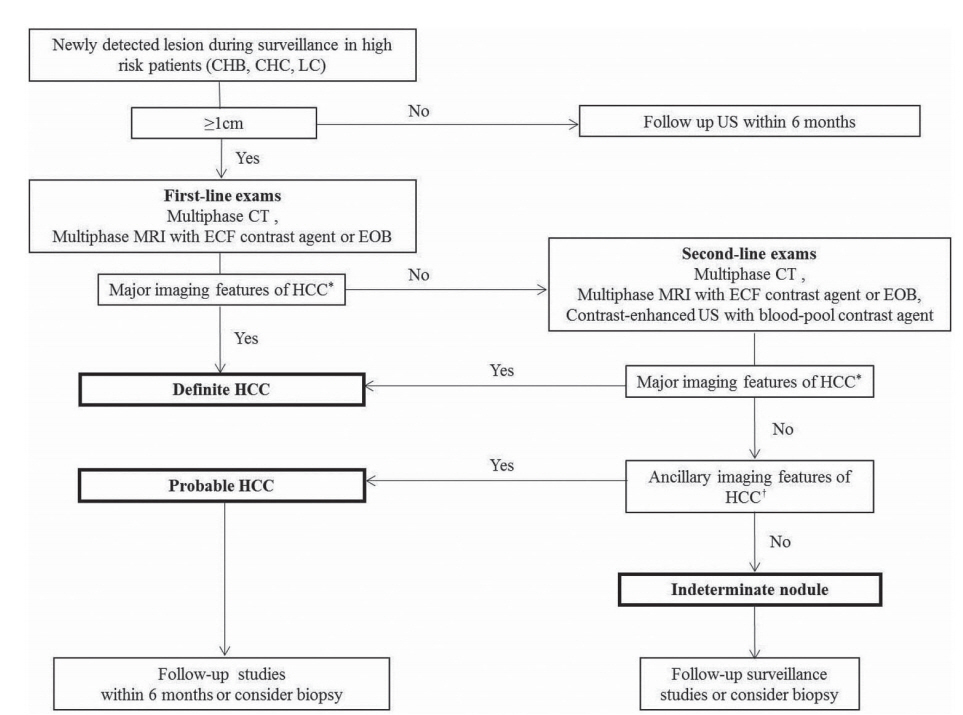
Figure 1.

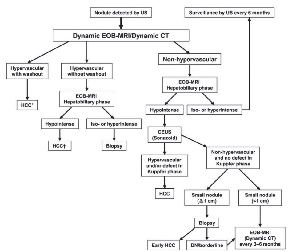
Figure 2.

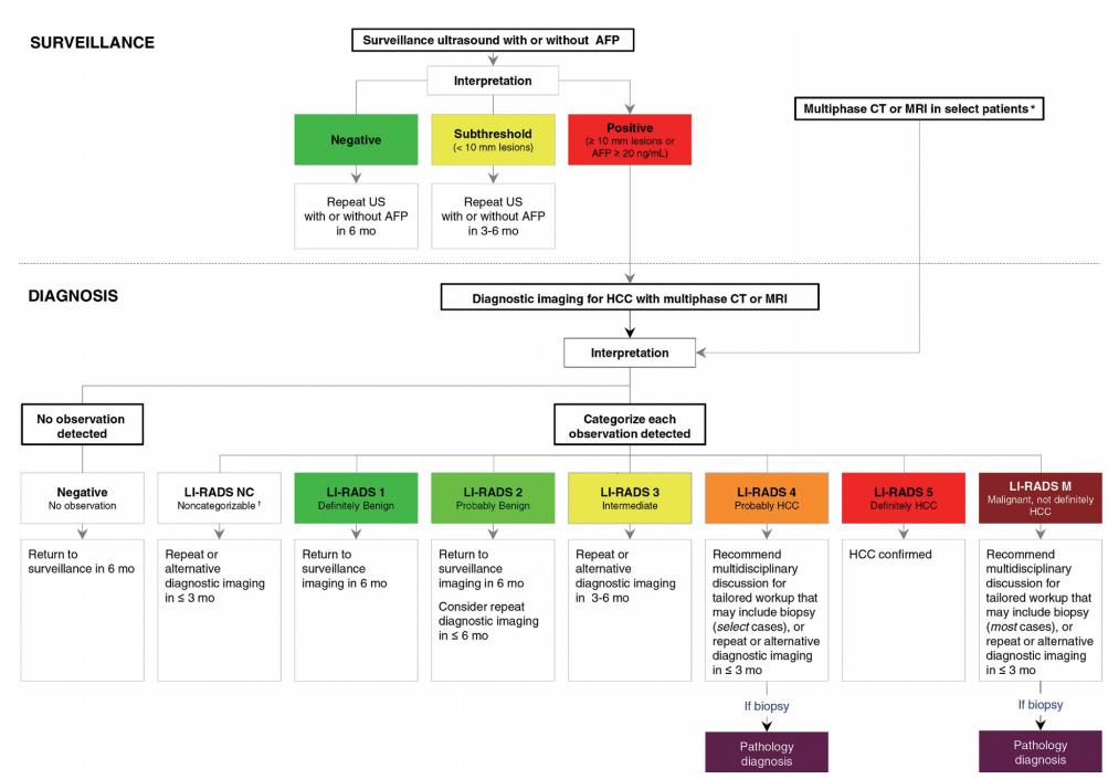
Figure 3.

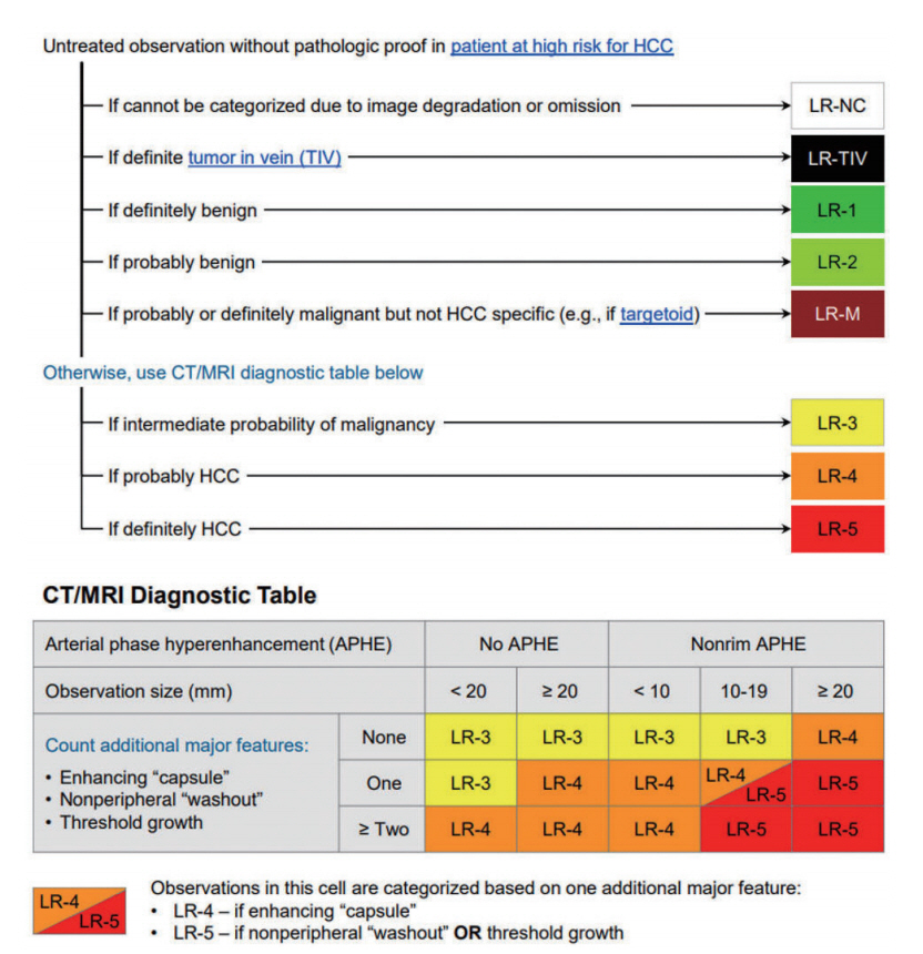
Figure 4.

Figure 5.

Table 1.
| Organizations | KLCA-NCC 2018 [13] | APASL 2017 [8] | AASLD 2018 [11] | LI-RADS 2018 [10] | EASL 2018 [12] |
|---|---|---|---|---|---|
| Target population for surveillance | âĒ Cirrhotic patients with varying etiology | âĒ Cirrhotic patients with varying etiology (HBV, HCV, NASH, genetic hemochromatosis, PBC, alpha-1 antitrypsin deficiency) | âĒ Cirrhotic patients with varying etiology (hepatitis B, hepatitis C, primary biliary cirrhosis, genetic hemochromatosis, alpha-1-antitrypsin) | âĒ Cirrhotic patients with any etiology | âĒ Cirrhotic patients, Child-Pugh stage A and B |
| âĒ Chronic HBV or HCV carrier | âĒ Noncirrhotic HBV carriers (Asian men >40 y, Asian women >50 y, African/North American blacks, family history of HCC) | âĒ Cirrhotic patients, Child- Pugh stage C awaiting liver transplantation | |||
| âĒ Non-cirrhotic HBV patients (Asian men >40 y, Asian women >50 y, Africans >20 y; family history of HCC) | âĒ Hepatitis B carriers (Asian men >40 y, Asian women > 50 y, all cirrhotic HBV carriers, family history of HCC, African/North American blacks) | âĒ Defers to regional HCC clinical practice guidelines for additional indications in the absence of cirrhosis | âĒ Non-cirrhotic HBV patients at intermediate or high risk of HCC | ||
| âĒ Non-cirrhotic patients with F3 fibrosis, regardless of etiology may be considered based on individual risk assessment | |||||
| Screening and surveillance test | âĒ Ultrasound and AFP measurements every 6 mo | âĒ Ultrasound and AFP measurements every 6 mo | âĒ Ultrasound with/without AFP every 6 mo | âĒ Ultrasound every 6 mo | âĒ Ultrasound every 6 mo |
| âĒ CT or MRI may be used in select patients with a high likelihood of having an inadequate ultrasound | âĒ CT or MRI may be utilized in select patients with a high likelihood of having an inadequate US or with performed but inadequate US | âĒ CT or MRI for patients on waiting list for liver transplantation and when obesity, intestinal gas, and chest wall deformity prevent adequate ultrasound assessment | |||
| âĒ Ultrasound <4 mo interval when a nodule of <1 cm has been detected during surveillance |
KLCA-NCC, Korean Liver Cancer Association-National Cancer Center; APASL, Asian-Pacific Association for the Study of the Liver; AASLD, Association for the Study of Liver Diseases; LI-RADS, Liver Imaging Reporting and Data System; EASL, European Association for the Study of the Liver; HBV, hepatitis B virus; HCV, hepatitis C virus; NASH, non alcoholic steatohepatitis; PBC, primary biliary cholangitis; y, years; HCC, hepatocellular carcinoma; AFP, alpha-fetoprotein; CT, computed tomography; MRI, magnetic resonance imaging; US, ultrasonography; mo, months.
Table 2.
| Organizations | KLCA-NCC 2018 [13] | APASL 2017 [8] | AASLD 2018 [11] | LI-RADS 2018 [10] | EASL 2018 [12] | |||
|---|---|---|---|---|---|---|---|---|
| Target population for diagnostic imaging | Patients at risk for HCC with âĨ1 cm nodule on screening/surveillance ultrasound | Patients at risk for HCC with positive screening/surveillance test or clinical suspicion of HCC | Patients at risk for HCC with abnormal results on screening/surveillance test or with clinical suspicions of HCC | âĒ Adult patients with cirrhosis of any cause except vascular disorder or congenital hepatic fibrosis | Patients at risk for HCC with âĨ1 cm nodule on screening/surveillance ultrasound | |||
| âĒ Patients with chronic hepatitis B with or without cirrhosis | ||||||||
| âĒ Patients with current or prior HCC with or without cirrhosis | ||||||||
| âĒ Adult liver transplantation candidates and liver transplant recipients | ||||||||
| Primary imaging modality | CT, MRI using ECCM or HBA | CT, MRI using ECCM or HBA | CT, MRI using ECCM or HBA | âĒ CT, MRI using ECCM or HBA | CT, MRI using ECCM or HBA | |||
| âĒ CEUS | ||||||||
| Secondary imaging modality | CEUS | CEUS (Sonazoid) | None | None | CEUS | |||
| Phases accepted for washout appearance | âĒ ECA: PVP or DP | âĒ ECA: PVP or DP | âĒ ECA: PVP or DP | âĒ ECA: PVP or DP | âĒ ECA: PVP or DP | |||
| âĒ HBA: PVP, DP or hypointensity on HBP | âĒ HBA: PVP | âĒ HBA: PVP | âĒ HBA: PVP | âĒ HBA: PVP | ||||
| Imaging criteria for arterial phase hyperenhancing HCC | âĒ Nodule size >1 cm | âĒ Regardless of size: | Defers to LI-RADS 5 category | âĒ Definitely HCC (LR-5) definition on CT/MRI: | âĒ Nodule size >1 cm | |||
| âĒ APHE and | âĒ APHE and | 1) Nodule size âĨ20 mm | âĒ APHE | |||||
| âĒ Washout | âĒ Washout on PVP or hypointensity on HBP | APHE and one or more of following: | âĒ Washout | |||||
| -Nonperipheral âwashoutâ | ||||||||
| -Enhancing capsule | ||||||||
| -Threshold growth | ||||||||
| 2) Nodule size 10-19 mm | ||||||||
| (1) APHE and | ||||||||
| -Nonperipheral âwashoutâ | ||||||||
| or | ||||||||
| -Threshold growth | ||||||||
| (2) APHE and two or more of the following: | ||||||||
| -Nonperipheral âwashoutâ | ||||||||
| -Enhancing capsule | ||||||||
| -Threshold growth | ||||||||
| âĒ Definitely HCC (LR-5) definition on CEUS: | ||||||||
| -Nodule size âĨ10 mm | ||||||||
| -APHE and | ||||||||
| -Late (>60 s) and mild washout | ||||||||
| Imaging criteria for arterial phase hypo- or isoenhancing HCC | None | Yes | None | None | None | |||
| Imaging criteria for arterial phase hypo- or isoenhancing PROBABLE HCC | Yes | None (but definite HCC diagnosis is possible) | Yes | Yes | None | |||
| Imaging criteria for subcentimeter size HCC | None | Yes | None | None | None | |||
| Exclusion criteria | Yes | None | None | None | None | |||
| When HBA is used, | ||||||||
| -T2 bright SI | ||||||||
| -Targetoid appearance in the DWI orCE-T1WI | ||||||||
| Imaging criteria for HCC tumor in vein | None | None | None | LR-TIV (unequivocal enhancing soft tissue TIV, regardless of visualization of a parenchymal mass) | None | |||
| Ancillary features | Yes | None | Yes | Yes | None | |||
| -To diagnose PROBABLE HCC | -Upgrading (up to LR-4) | |||||||
| -Downgrading | ||||||||
| Categories | âĒ Arterial hyperenhancing HCC | âĒ Arterial hyperenhancing HCC | âĒ Definitely benign (LR-1) | âĒ Definitely benign (LR-1) | Arterial hyperenhancing HCC | |||
| âĒ Probably benign (LR-2) | âĒ Probably benign (LR-2) | |||||||
| âĒ PROBABLE HCC | âĒ Arterial hypo- isoenhancing HCC | âĒ Intermediate (LR-3) | âĒ Intermediate probability of malignancy (LR-3) | |||||
| âĒ Probably HCC (LR-4) | âĒ Probably HCC (LR-4) | |||||||
| âĒ Definitely HCC (LR-5) | âĒ Definitely HCC (LR-5) | |||||||
| âĒ Malignant, not definitely HCC (LR-M) | âĒ Definite tumor in vein (LR-TIV) | |||||||
| âĒ Probably or definitely malignant but not HCC specific (LR-M) | ||||||||
| Staging | mUICC | Do not specify any staging system (multidisciplinary discussion is required) | BCLC | Radiologic T-staging | BCLC | |||
KLCA-NCC, Korean Liver Cancer Association-National Cancer Center; APASL, Asian-Pacific Association for the Study of the Liver; AASLD, Association for the Study of Liver Diseases; LI-RADS, Liver Imaging Reporting and Data System; EASL, European Association for the Study of the Liver; HCC, hepatocellular carcinoma; CT, computed tomography; MRI, magnetic resonance imaging; ECCM, extracellular contrast media; HBA, hepatobiliary contrast agent; CEUS, contrast enhanced ultrasound; ECA, extracellular contrast agents; PVP, portal venous phase; DP, delayed phase; HBP, hepatobiliary phase; APHE, arterial phase hyperenhancement; SI, signal intensity; DWI, diffusion weighted image; CE, contrast enhance; TIV, tumor in vein; mUICC, Modified Union for International Cancer Control; BCLC, Barcelona Clinic Liver Cancer.
Abbreviations
REFERENCES
- TOOLS
-
METRICS

- ORCID iDs
-
Jeong Min Lee

https://orcid.org/0000-0003-0561-8777 - Related articles
-
The prime time for management of hepatocellular carcinoma in Hong Kong2023 April;29(2)


 PDF Links
 PubReader
 ePub Link
 Full text via DOI
 Full text via PMC
 Download Citation
 Print
Print


