Clin Mol Hepatol > Volume 29(3); 2023 > Article
Park, Chung, Lee, Shin, Hur, Cho, Park, Youk, Lee, Lee, Yu, Kim, Yoon, Kim, and Lee: Treated chronic hepatitis B is a good prognostic factor of diffuse large B-cell lymphoma

ABSTRACT

Background/Aims

Chronic hepatitis B (CHB) is a risk factor for non-Hodgkin lymphoma (NHL). Our recent study suggested that antiviral treatment may reduce the incidence of NHL in CHB patients. This study compared the prognoses of hepatitis B virus (HBV)-associated diffuse large B-cell lymphoma (DLBCL) patients receiving antiviral treatment and HBV-unassociated DLBCL patients.

Methods

This study comprised 928 DLBCL patients who were treated with rituximab, cyclophosphamide, doxorubicin, vincristine, and prednisone (R-CHOP) at two referral centers in Korea. All patients with CHB received antiviral treatment. Time-to-progression (TTP) and overall survival (OS) were the primary and secondary endpoints, respectively.

Results

Among the 928 patients in this study, 82 were hepatitis B surface antigen (HBsAg)-positive (the CHB group) and 846 were HBsAg-negative (the non-CHB group). The median follow-up time was 50.5 months (interquartile range [IQR]=25.6–69.7 months). Multivariable analyses showed longer TTP in the CHB group than the non-CHB group both before inverse probability of treatment weighting (IPTW; adjusted hazard ratio [aHR]=0.49, 95% confidence interval [CI]=0.29–0.82, P=0.007) and after IPTW (aHR=0.42, 95% CI=0.26–0.70, P<0.001). The CHB group also had a longer OS than the non-CHB group both before IPTW (HR=0.55, 95% CI=0.33–0.92, log-rank P=0.02) and after IPTW (HR=0.53, 95% CI=0.32–0.99, log-rank P=0.02). Although liver-related deaths did not occur in the non-CHB group, two deaths occurred in the CHB group due to hepatocellular carcinoma and acute liver failure, respectively.

Conclusions

Our findings indicate that HBV-associated DLBCL patients receiving antiviral treatment have significantly longer TTP and OS after R-CHOP treatment than HBV-unassociated DLBCL patients.

Graphical Abstract

INTRODUCTION

Hepatitis B virus (HBV) infection is the most prevalent chronic viral infection globally and is associated with the development of liver disease, including hepatocellular carcinoma (HCC) [1-3]. HBV infection is also a recognized risk factor for non-Hodgkin lymphoma (NHL) [4,5]. Diffuse large B-cell lymphoma (DLBCL) is a common type of NHL, and a significant association between chronic hepatitis B (CHB) and DLBCL development has been reported [6]. Consequently, CHB is considered a major risk factor for DLBCL [4,5,7].
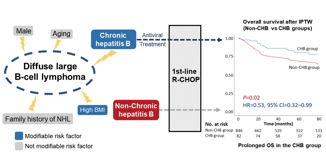
In our recent population-based study, CHB patients had an increased risk of NHL, and antiviral therapy was suggested to attenuate the risk of NHL to be comparable to that of non-CHB subjects [8]. This result supports the hypothesis that active HBV replication could contribute to NHL development in CHB patients. However, whether antiviral treatment decreases the disease progression or recurrence of HBV-associated NHL or DLBCL is debatable [4,7,9]. Several risk factors for DLBCL have been identified, including B-cell-activating autoimmune disease, hepatitis C virus (HCV) positivity, first-degree familial history of NHL, high body mass index, and HBV infection [10,11]. Many of these risk factors are not modifiable; however, HBV infection is modifiable through antiviral treatment. Thus, we hypothesized that the disease course of HBV-associated DLBCL patients may be improved with antiviral therapies, similar to patients with HBV-unassociated DLBCL resulting from other non-modifiable risk factors.
In the present study, the prognoses of HBV-associated DLBCL patients receiving antiviral treatment and of HBV-unassociated DLBCL patients were compared. All patients received rituximab, cyclophosphamide, doxorubicin, vincristine, and prednisone (R-CHOP) as first-line chemotherapy.

MATERIALS AND METHODS

Study population

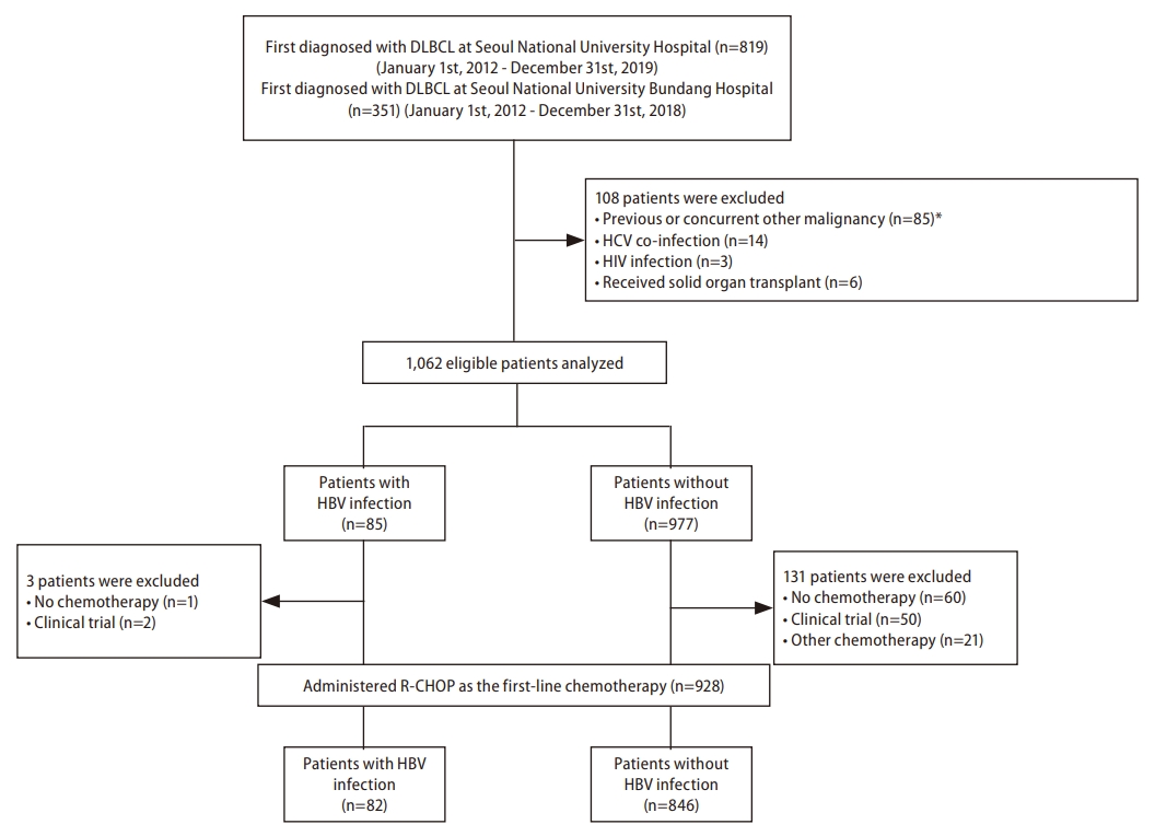
We retrospectively included patients who were first diagnosed with DLBCL and received R-CHOP treatment as first-line chemotherapy at Seoul National University Hospital (SNUH; Seoul, Korea) between January 1, 2012, and December 31, 2019, or at Seoul National University Bundang Hospital (SNUBH; Seongnam, Korea) between January 1, 2012, and December 31, 2018. DLBCL was diagnosed histologically according to the 2008 and 2016 World Health Organization classification of tumors of hematopoietic and lymphoid tissues [12,13]. The cell of origin was defined as a germinal center B cell (GCB) or non-GCB subtype based on the Hans algorithm [14]. We excluded patients who were diagnosed with other malignancies within 5 years before DLBCL diagnosis, had other, concurrent viral infections (i.e., HCV, hepatitis D virus, or human immunodeficiency virus [HIV]) or who had undergone organ transplantation. Based on the presence of hepatitis B surface antigen (HBsAg) or HBV DNA, patients were classified into either the CHB group or the non-CHB group (Fig. 1).
All DLBCL patients underwent routine serology screening including HBsAg and anti-HBV core antibody (anti-HBc) before chemotherapy to determine the risk of HBV reactivation or flare [15]. HBsAg and anti-HBc were examined using the ARCHITECT i2000SR (Abbott Laboratories, Abbott Park, IL, USA) and the Alinity i system (Abbott Laboratories), respectively. CHB patients who were already being treated with oral nucleos(t)ide analogues (NAs) continued antiviral treatment. All untreated CHB patients positive for either HBsAg or anti-HBc started antiviral treatment with an NA before initiation of rituximab according to the CHB treatment guidelines [16-19]. Prophylactic antiviral agents were discontinued 12 months after completion of R-CHOP treatment if blood tests showed continuous HBV suppression. In cases of DLBCL recurrence requiring subsequent treatment, the prophylactic antiviral agents were resumed if indications were met. In addition, therapeutic antivirals were administered if a patient experienced HBV reactivation and a hepatitis flare-up following cessation of prophylactic antivirals [17,20].
This study conformed to the ethical guidelines of the World Medical Association Declaration of Helsinki and was approved by the Institutional Review Board of Seoul National University Hospital (IRB No. H-2205-176-1329). The requirement for informed consent from patients was waived due to the retrospective nature of the study.

Definitions and assessments

The Ann Arbor and Lugano classifications were used for DLBCL staging [21,22]. Computed tomography (CT) and positron emission tomography-CT (PET-CT) were conducted to evaluate the extent of lesions at baseline. Bone marrow biopsy was performed to confirm bone marrow involvement of DLBCL. Performance status was presented as Eastern Cooperative Oncology Group scores. The International Prognostic Index (IPI) was used to predict the outcomes for individuals receiving rituximab in combination with chemotherapy [23]. CT or PET-CT was performed every two or three cycles of R-CHOP chemotherapy to evaluate the treatment response. Additional descriptions of Ann Arbor classification and IPI risk are provided in Supplementary Tables 1 and 2, respectively. The diagnostic criteria for cirrhosis are described in Supplementary Methods.

Outcomes and follow-up

The primary endpoint was time-to-progression (TTP) of DLBCL. The secondary endpoints were tumor response, progression-free survival (PFS), and overall survival (OS). The treatment response of NHL was assessed according to the Lugano criteria as complete remission (CR), partial response, stable disease, or progressive disease [21]. The index date was defined as the date on which the patient started R-CHOP treatment as first-line chemotherapy. Patients were censored at the last date of follow-up, the date of death, or the data cut-off date (i.e., April 30, 2022), whichever came first.
After each patient completed six cycles of R-CHOP, CT or PET-CT was performed to evaluate the treatment response, and a follow-up CT or PET-CT was performed every 3 months for 1 year after the last round of chemotherapy. An annual imaging follow-up (CT or PET-CT) was performed after patients maintained CR for 1 year. When a patient experienced symptoms of disease progression, unscheduled diagnostic studies including CT or PET-CT were performed according to the decision of attending physicians.
Statistical analysis Categorical variables were presented as frequencies (%) and continuous variables as medians and interquartile ranges (IQRs). Pearson’s χ2 test or Fisher’s exact test was used to compare categorical variables, and Student’s t-test or Mann–Whitney U test was used to compare continuous variables. The Holm–Bonferroni method was used to control for Type I error resulting from multiple comparisons (Supplementary Methods) [24]. Inverse probability of treatment weighting (IPTW) [25] was used to reduce potential confounding. Age, sex, Ann Arbor classification, IPI risk, GCB subtype, lactate dehydrogenase level, presence of an extranodal lesion, bone marrow involvement, and cirrhosis were included in the propensity score (PS) calculation. The Kaplan–Meier estimator and log-rank test were used to determine TTP, PFS, and OS. For risk factor analysis, factors found significant in univariable analysis or those with a low degree of collinearity were subjected to multivariable analysis using Cox regression analysis. P-values for interactions were used to evaluate the differential effect of current HBV infection on DLBCL clinical outcomes based on subgroups [26]. All statistical analyses were performed using R version 4.2.0 (R Foundation for Statistical Computing, Vienna, Austria). P-values <0.05 were considered statistically significant.

RESULTS

Baseline characteristics

The median follow-up duration was 50.5 months (IQR=25.6–69.7 months). The baseline characteristics of the study population are presented in Table 1. A total of 928 patients was included in this study; 82 had evidence of current CHB infection and were classified as the CHB group; the remaining 846 patients comprised the non-CHB group. After IPTW, the baseline characteristics were generally well balanced between the study groups (Table 1).

Comparison of outcomes between the CHB and non-CHB groups

The CHB and non-CHB groups had similar objective response rates (91.5% vs. 91.0%, P=1.00); however, the non-CHB group showed a greater proportion of SD or PD (1.2% vs. 6.5%, P=0.046; Supplementary Table 3).
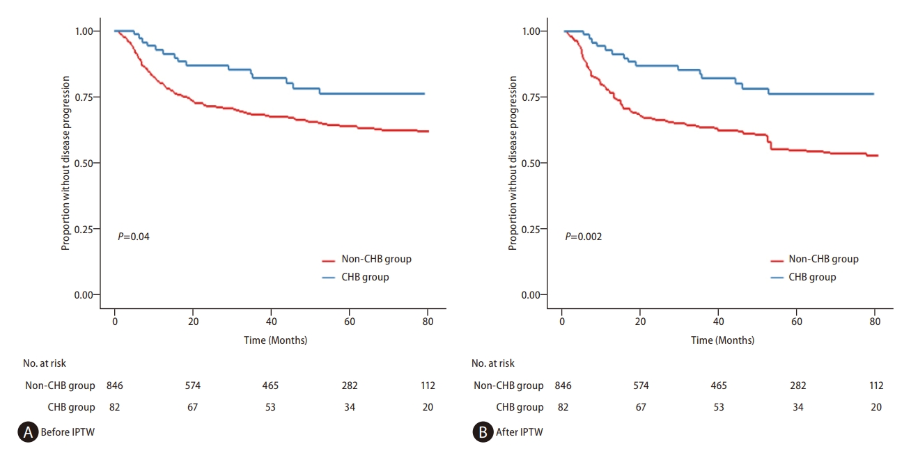
During the study period, 269 patients (29.0%) experienced tumor recurrence or DLBCL progression: 16 patients (19.5%) in the CHB group and 253 patients (29.9%) in the non-CHB group. TTP was significantly longer in the CHB group before IPTW (hazard ratio [HR]=0.58, 95% confidence interval [CI]=0.35–0.97, log-rank P=0.04) and remained significantly longer after IPTW (HR=0.49, 95% CI=0.29–0.82, log-rank P=0.002; Fig. 2 and Table 2). Multivariable analyses also showed longer TTP in the CHB group than the non-CHB group, both before IPTW (adjusted HR [aHR]=0.49, 95% CI=0.29–0.82, P=0.007) and after IPTW (aHR=0.42, 95% CI=0.26–0.70, P<0.001; Table 2). All the above results were statistically significant after the Holm–Bonferroni correction for multiple comparisons (Supplementary Results). Separate analyses of the SNUH and SNUBH subcohorts yielded the same results (Supplementary Table 4). TTP was significantly longer in the CHB group after IPTW in both SNUH (HR=0.51, 95% CI=0.29–0.92, log-rank P=0.03; Supplementary Fig. 1) and SNUBH subcohorts (HR=0.40, 95% CI=0.10–0.94, log-rank P=0.04; Supplementary Fig. 2). In the subgroup analyses, the difference in TTP between CHB and non-CHB groups was influenced by the presence of liver cirrhosis, bone marrow involvement, IPI risk group, and GCB subtype (Supplementary Table 5A). Subgroup analyses showed no variables affecting the difference in TTP between the CHB and non-CHB groups in the SNUH subcohort; however, in the SNUBH subcohort, elevated LDH affected the difference (Supplementary Table 5B, C). Difference was not observed in TTP based on hepatitis B e antigen status in CHB patients before IPTW (HR=0.58, 95% CI=0.07–4.43, log-rank P=0.59) and after IPTW (HR=0.63, 95% CI=0.11–7.50, log-rank P=0.93). A low IPI risk and an early Ann Arbor stage were associated with a significantly longer TTP in the CHB and non-CHB groups both before and after IPTW (Supplementary Figs. 3 and 4).
Univariable analyses showed a significant difference in PFS between the CHB and non-CHB groups both before IPTW (HR=0.61, 95% CI=0.40–0.94, log-rank P=0.03) and after IPTW (HR=0.53, 95% CI=0.34–0.82, log-rank P=0.004; Supplementary Fig. 5). Multivariable analyses also showed significantly longer PFS in the CHB group than the non-CHB group both before IPTW (aHR=0.56, 95% CI=0.37–0.87, P=0.01) and after IPTW (aHR=0.50, 95% CI=0.32–0.77, P=0.002; Supplementary Table 6). A marginal but not significant difference in PFS was observed between the two groups after IPTW in both SNUH (HR=0.63, 95% CI=0.39–1.02, log-rank P=0.06; Supplementary Fig. 6) and SNUBH subcohorts (HR=0.32, 95% CI=0.10–1.04, log-rank P=0.06; Supplementary Fig. 7).
In the CHB group, OS was also significantly longer than in the non-CHB group both before IPTW (HR=0.55, 95% CI=0.33–0.92, log-rank P=0.02; Fig. 3A) and after IPTW (HR=0.53, 95% CI=0.32–0.99, log-rank P=0.02; Fig. 3B). Multivariable analyses indicated that the CHB group was independently associated with a longer OS than the non-CHB group both before IPTW (aHR=0.55, 95% CI=0.33–0.93, P=0.03; Table 3A) and after IPTW (aHR=0.49, 95% CI=0.29–0.81, P=0.007; Table 3B). Regarding liver-related deaths, 2 patients in the CHB group who had cirrhosis at baseline died: 1 due to newly developed HCC and 1 due to acute liver failure. In the sensitivity analyses that excluded these 2 patients and involved reanalysis of the data, the results for TTP remained consistent (Supplementary Results). In the non-CHB group, liver-related death did not occur, and the risk of liver failure was not significantly different between the two groups (7.1% vs. 0.0%, P=0.07; Supplementary Table 7). Non-liver-related deaths occurred less frequently in the CHB group than in the non-CHB group after IPTW (HR=0.48, 95% CI=0.27–0.88, log-rank P=0.02; Supplementary Fig. 8). Although OS tended to be longer in the CHB group, difference was not observed between the two groups in either SNUH (Supplementary Fig. 9) or SNUBH subcohort (Supplementary Fig. 10) before and after IPTW. Lower IPI risk and early Ann Arbor stage were associated with significantly longer TTP in the CHB and non-CHB groups both before and after IPTW. A low IPI risk and an early Ann Arbor stage were also associated with significantly longer PFS and OS in the CHB and non-CHB groups both before and after IPTW (Supplementary Figs. 1114).

Comparisons of TTP among the CHB, resolved-HBV, and never-HBV groups

The non-CHB group was further subclassified into resolved-HBV (HBsAg-negative but anti-HBc-positive) or never-HBV (HBsAg-negative and anti-HBc-negative) groups. Within the non-CHB group, the resolved-HBV group was older, had a larger proportion of patients with cirrhosis, and had higher IPI risk scores than the never-HBV group (Supplementary Table 8). Significant difference was not observed in TTP before IPTW between the CHB and never-HBV groups (HR=0.65, 95% CI=0.39–1.09, log-rank P=0.10). However, after IPTW, the CHB group had a longer TTP than the never-HBV group (HR=0.52, 95% CI=0.31–0.88, log-rank P=0.01). Both before and after IPTW, significant differences were observed in TTP between the CHB and resolved-HBV groups before IPTW (HR=0.47, 95% CI=0.28–0.80, log-rank P=0.005) and after IPTW (HR=0.43, 95% CI=0.25–0.75, log-rank P=0.003; Supplementary Fig. 15). TTP was similar between never-HBV and resolved-HBV patients after IPTW (HR=1.12, 95% CI=0.88–1.53, log-rank P=0.30).

DISCUSSION

In this study, we examined the clinical outcomes of DLBCL patients treated with R-CHOP based on the HBV infection status. After baseline characteristics were balanced with IPTW, patients with CHB had a 51–58% lower risk of progression. In addition, the CHB group showed longer PFS and OS than the non-CHB group. When the non-CHB group was subdivided into the resolved-HBV or never-HBV groups, the CHB group showed significantly longer TTP than both the re-solved-HBV and never-HBV groups; however, significant difference was not observed in TTP between the resolved-HBV and never-HBV groups. In subgroup analysis, the results were comparable between SNUH and SNUBH subcohorts.
Prior to the development of antivirals, many HBsAg-positive lymphoma patients died of severe hepatitis caused by HBV reactivation [15]. The risk of HBV reactivation during chemotherapy for lymphoma is significantly reduced by antiviral prophylaxis [27]. Consequently, current practice guidelines advocate antiviral prophylaxis for patients being treated with chemotherapeutic agents, particularly rituximab [16,18]. Clinical and epidemiological evidence further supports an association of HBV with DLBCL development [6,8]; however, the extrahepatic effect of HBV suppression by NAs on HBV-associated DLBCL remained unknown. Our study shows that current HBV infection is a favorable prognostic factor for DLBCL patients in the era of antiviral prophylaxis.
Multiple mechanisms may explain why antiviral treatment can prolong TTP. First, reducing HBV viral load inhibits HBV-driven antigen stimulation of B cells, inhibiting lymphoma progression or recurrence [28]. In the majority of HBsAg-positive DLBCL patients, the third complementarity-determining region of heavy- or light-chain sequences displayed high homology to antibody-targeting HBsAg [28], indicating that these DLBCL cells originated from B cells reactive to HBV due to chronic HBV antigen stimulation. If persistent HBV-driven antigen stimulation is a primary cause of DLBCL development, then blocking antigen stimulation can inhibit one of the major proliferative and anti-apoptotic signals in HBV-associated DLBCL. Second, decreasing the HBV viral load reduces the HBV-mediated apolipoprotein B mRNA-editing catalytic polypeptide (APOBEC)-induced mutagenesis [29,30]. APOBEC3 cytidine deaminases are essential for regulating gene expression, innate immunity, and suppressing HBV infection. Aberrant expression during chronic viral infections or inflammation can cause the development of cancer [31-33]. APOBEC3 genes are significantly increased in CHB patients and may induce extrahepatic malignancy through the migration of peripheral blood mononuclear cells or lymphoid cells with upregulated APOBEC3 expression in response to HBV infection [34,35]. In a gene expression study on DLBCL patients, an association was found between APOBEC3 genes and poor prognosis, indicating these genes may contribute to the development of DLBCL [36]. Therefore, reducing HBV viral load with antiviral agents can decrease APOBEC3 expression and potentially lead to a reduction in cancer development [37]. Third, both the innate and adaptive immune systems are dysfunctional in patients with a high HBV viral load, resulting in decreased immune surveillance [38]. However, antiviral treatment restores immune surveillance of malignancy, reducing the likelihood of lymphoma recurrence [39,40].
In the subgroup analysis, the CHB group of cirrhotic patients tended to have a shorter TTP than the non-CHB group. Furthermore, the cirrhotic CHB patients did not receive significantly lower doses of chemotherapeutic agents than the non-cirrhotic patients (data not shown). However, because the progression of cirrhosis is reportedly associated with persistent active viral replication, cirrhotic HBV patients are expected to be exposed to higher viral loads than non-cirrhotic patients [41]. Consequently, cirrhotic patients may have a higher risk of prolonged HBV-mediated APOBEC-induced mutagenesis, leading to an increased mutation burden of DLBCL, which is associated with a poor prognosis. Immune dysfunction in cirrhotic patients may possibly contribute to DLBCL recurrence by reducing immune surveillance; however, further research on the underlying mechanisms is warranted [42].
The results of the present study revealed that patients with current HBV infection (i.e., HBsAg-positive) and patients with resolved HBV infection (i.e., HBsAg-negative but anti-HBcpositive) showed distinct clinical trajectories. This finding indicates that current HBV infection is a risk factor for development and recurrence of DLBCL. According to previous studies, infected HBV continuously stimulates B cells and facilitates mutagenesis, both of which lead to DLBCL development [28,43]. However, neither chronic HBV antigen stimulation nor HBV-mediated mutagenesis occurs in patients with resolved HBV infection. Therefore, antiviral therapy may have limited chemopreventive effects in patients where DLBCL development occurs through alternate pathways.
Off-target effects of antivirals may also be insufficient to explain their preventive effect on DLBCL progression or recurrence. For example, tenofovir is a potent antiviral that blocks HBV viral replication and induces interferon lambda 3 (IFN-λ3) production, exhibiting additional unanticipated (i.e., off-target) pharmacological effects [44]. IFN-λ has antitumor effects by inducing cell cycle arrest and causing apoptosis [45,46]. If this off-target effect of antivirals plays a role in preventing lymphoma progression or recurrence, patients with resolved HBV should show a trend either similar to that of CHB patients or superior to never-HBV patients. However, resolved-HBV patients showed a trend similar to never-HBV patients. Therefore, genetic factors, such as mutational load, appear to be more important in the progression of lymphoma than the off-target effects of antivirals. Studies evaluating the genetic differences between DLBCL patients with resolved HBV and those with current HBV are warranted.
In addition, significant differences were observed in OS between the CHB and non-CHB groups both before and after IPTW. Although antiviral treatment reduces HBV reactivation in CHB patients, the possibility of liver-related death is higher in these patients than in non-CHB patients [47]. Even after antiviral treatment, CHB patients have a greater risk of HCC6 and drug-induced liver injury [48,49] than non-CHB patients, both of which could lead to mortality. Despite these factors, the significantly extended OS in the CHB group compared with the non-CHB group indicates that, in HBV-associated DLBCL, antiviral agents suppress HBV replication and have a synergetic effect with R-CHOP, contributing to prolonged survival.
The present study had several limitations. First, because this was a retrospective study, selection bias may have occurred. However, we conducted a multicenter study and applied multiple statistical methods, including IPTW and multivariable analysis, to overcome this potential bias. Second, the population was restricted to DLBCL patients initially treated with R-CHOP. Patients with HBV-associated DLBCL who underwent chemotherapies other than R-CHOP may experience distinct clinical outcomes. However, by restricting the study population to only these patients, the influence of chemotherapeutic regimens on DLBCL was minimized, focusing on HBV viral load and clinical outcomes.
In conclusion, HBV infection was a favorable prognostic factor associated with a prolonged TTP, PFS, and OS in HBV-related DLBCL patients who received antiviral medication prior to R-CHOP chemotherapy.

ACKNOWLEDGMENTS

This work wassupported by National IT Industry Promotion Agency grant funded by the Korea Ministry of Science and ICT [No. S0252-21-1001), Liver Research Foundation of Korea as part of Bio Future Strategies Research Project.

FOOTNOTES

Authors’ contribution
The corresponding authors (Tae Min Kim and Jeong-Hoon Lee) had full access to all the data in the study and take responsibility for the integrity of the data and the accuracy of the data analysis. Specific author contributions: Conceptualzation and Methodology, Jeayeon Park, Sung Won Chung, and Jeong-Hoon Lee; Formal Analysis, Jeayeon Park and Sung Won Chung; Data Curation and Investigation, Jeayeon Park, Sung Wong Chung, Hyunjae Shin, Moon Haeng Hur, and Heejin Cho; Resources, Min Kyung Park, Jeonghwan Youk, Ji Yun Lee, Yun Bin Lee, Jeong-Ok Lee, Su Jong Yu, Yoon Jun Kim, Jung-Hwan Yoon, Tae Min Kim, and Jeong-Hoon Lee; Writing – Original Draft Preparation, Jeayeon Park and Sung Won Chung; Writing – Review & Editing, Jeayeon Park, Sung Won Chung, Yun Bin Lee, Tae Min Kim, and Jeong-Hoon Lee; Visualization, Jeayeon Park and Sung Won Chung; Supervision, Tae Min Kim and Jeong-Hoon Lee; Project Administration, Tae Min Kim and Jeong-Hoon Lee; Approval of the final article: all authors.
Conflicts of Interest
Dr. Yun Bin Lee reportsreceiving research grantsfrom Samjin Pharmaceuticals and Yuhan Pharmaceuticals; Dr. Su Jong Yu reports receiving research grants from Yuhan Pharmaceuticals and Daewoong Pharmaceuticals; Dr. Yoon Jun Kim reports receiving research grants from Yuhan Pharmaceuticals, AstraZeneca, Boston Scientific, Daewoong Pharmaceuticals, and Samjin Pharmaceuticals and lecture fees from Bayer HealthCare Pharmaceuticals, Gilead Science, MSD Korea, Yuhan Pharmaceuticals, Samil Pharmaceuticals, CJ Pharmaceuticals, Bukwang Pharmaceuticals, and Handok Pharmaceuticals; Dr. Jung-Hwan Yoon reports receiving research grants from Dicerna Pharmaceuticals, Roche, GlaxoSmithKline, AstraZeneca, Daewoong Pharmaceuticals, and Hanmi Pharmaceuticals; Dr. Tae Min Kim reports receiving research grants from AstraZeneca-Korea Health Industry Development Institute and honoraria or advisory role in AstraZeneca, Boryung, F. Hoffmann-La Roche Ltd/Genentech, Inc, Janssen, Novartis, Regeneron, Sanofi, Takeda, and Yuhan; Dr. Jeong-Hoon Lee reports receiving research grants from Yuhan Pharmaceuticals, and lecture fee from GreenCross Cell, Daewoong Pharmaceuticals, and Gilead Korea. No other potential conflict of interest relevant to this articlewasreported.

SUPPLEMENTAL MATERIAL

Supplementary material is available at Clinical and Molecular Hepatology website (http://www.e-cmh.org).
Supplementary Methods
cmh-2023-0057-Supplementary-1.pdf
Supplementary Results
cmh-2023-0057-Supplementary-2.pdf
Supplementary References
cmh-2023-0057-Supplementary-3.pdf
Supplementary Table 1.
Ann Arbor classification
cmh-2023-0057-Supplementary-Table-1.pdf
Supplementary Table 2.
International Prognostic Index for DLBCL
cmh-2023-0057-Supplementary-Table-2.pdf
Supplementary Table 3.
Treatment responses of DLBCL before and after IPTW
cmh-2023-0057-Supplementary-Table-3.pdf
Supplementary Table 4.
Baseline characteristics of (A) SNUH and (B) SNUBH subcohorts (A) SNUH subcohort
cmh-2023-0057-Supplementary-Table-4.pdf
Supplementary Table 5.
Cox regression analysis of subgroups associated with TTP of (A) entire cohort, (B) SNUH subcohort, and (C) SNUBH subcohort between the CHB and non-CHB groups af ter IPTW
cmh-2023-0057-Supplementary-Table-5.pdf
Supplementary Table 6.
Cox regression analysis of factors associated PFS (A) before and (B) after IPTW
cmh-2023-0057-Supplementary-Table-6.pdf
Supplementary Table 7.
Causes of death
cmh-2023-0057-Supplementary-Table-7.pdf
Supplementary Table 8.
Baseline characteristics of the resolved- and never-HBV groups before and after IPTW
cmh-2023-0057-Supplementary-Table-8.pdf
Supplementary Figure 1.
Kaplan–Meier estimates of TTP according to HBV infection status (A) before and (B) after IPTW in the SNUH subcohort. Propensity scores of inverse probability of treatment weighting were computed using age, sex, Ann Arbor classification, International Prognostic Index risk, germinal center B cell subtype, lactate dehydrogenase, presence of extranodal lesions, bone marrow involvement, and cirrhosis. CHB, chronic hepatitis B; HBV, hepatitis B virus; SNUH, Seoul National University Hospital; TTP, time-to-progression.
cmh-2023-0057-Supplementary-Fig-1.pdf
Supplementary Figure 2.
Kaplan–Meier estimates of TTP according to HBV infection status (A) before and (B) after IPTW in the SNUBH subcohort. Propensity scores of inverse probability of treatment weighting were computed using age, sex, Ann Arbor classification, International Prognostic Index risk, germinal center B cell subtype, lactate dehydrogenase, presence of extranodal lesions, bone marrow involvement, and cirrhosis. CHB, chronic hepatitis B; HBV, hepatitis B virus; SNUBH, Seoul National University Bundang Hospital; TTP, time-to-progression.
cmh-2023-0057-Supplementary-Fig-2.pdf
Supplementary Figure 3.
Kaplan–Meier estimates of TTP according to IPI risk and HBV infection status between the CHB and non-CHB groups (A) before and (B) after IPTW. Propensity scores of inverse probability of treatment weighting were computed using age, sex, Ann Arbor classification, IPI risk, germinal center B cell subtype, lactate dehydrogenase, presence of extranodal lesions, bone marrow involvement, and cirrhosis. CHB, chronic hepatitis B; HBV, hepatitis B virus; High-I, High-Intermediate; IPI, International Prognostic Index; Low-I, Low-Intermediate; TTP, time-to-progression.
cmh-2023-0057-Supplementary-Fig-3.pdf
Supplementary Figure 4.
Kaplan–Meier estimates of TTP according to Ann Arbor classification and HBV infection status between the CHB and non-CHB groups (A) before and (B) after IPTW. Propensity scores of inverse probability of treatment weighting were computed using age, sex, Ann Arbor classification, International Prognostic Index risk, germinal center B cell subtype, lactate dehydrogenase, presence of extranodal lesions, bone marrow involvement, and cirrhosis. CHB, chronic hepatitis B; HBV, hepatitis B virus; TTP, time-to-progression.
cmh-2023-0057-Supplementary-Fig-4.pdf
Supplementary Figure 5.
Kaplan–Meier estimates of PFS according to HBV infection status between the CHB and non-CHB groups (A) before and (B) after IPTW. Propensity scores of inverse probability of treatment weighting were computed using age, sex, Ann Arbor classification, International Prognostic Index risk, germinal center B cell subtype, lactate dehydrogenase, presence of extranodal lesions, bone marrow involvement, and cirrhosis. CHB, chronic hepatitis B; HBV, hepatitis B virus; PFS, progression-free survival.
cmh-2023-0057-Supplementary-Fig-5.pdf
Supplementary Figure 6.
Kaplan–Meier estimates of PFS according to HBV infection status (A) before and (B) after IPTW in the SNUH subcohort. Propensity scores of inverse probability of treatment weighting were computed using age, sex, Ann Arbor classification, International Prognostic Index risk, germinal center B cell subtype, lactate dehydrogenase, presence of extranodal lesions, bone marrow involvement, and cirrhosis. CHB, chronic hepatitis B; HBV, hepatitis B virus; PFS, progression-free survival; SNUH, Seoul National University Hospital.
cmh-2023-0057-Supplementary-Fig-6.pdf
Supplementary Figure 7.
Kaplan–Meier estimates of PFS according to HBV infection status (A) before and (B) after IPTW in the SNUBH subcohort. Propensity scores of inverse probability of treatment weighting were computed using age, sex, Ann Arbor classification, International Prognostic Index risk, germinal center B cell subtype, lactate dehydrogenase, presence of extranodal lesions, bone marrow involvement, and cirrhosis. Abbreviations: CHB, chronic hepatitis B; HBV, hepatitis B virus; PFS, progression-free survival; SNUBH, Seoul National University Bundang Hospital.
cmh-2023-0057-Supplementary-Fig-7.pdf
Supplementary Figure 8.
Cumulative risk of non-liver-related death according to HBV infection status (A) before and (B) after IPTW. Propensity scores of inverse probability of treatment weighting were computed using age, sex, Ann Arbor classification, International Prognostic Index risk, germinal center B cell subtype, lactate dehydrogenase, presence of extranodal lesions, bone marrow involvement, and cirrhosis. CHB, chronic hepatitis B; HBV, hepatitis B virus.
cmh-2023-0057-Supplementary-Fig-8.pdf
Supplementary Figure 9.
Kaplan–Meier estimates of OS according to HBV infection status (A) before and (B) after IPTW in the SNUH subcohort. Propensity scores of inverse probability of treatment weighting were computed using age, sex, Ann Arbor classification, International Prognostic Index risk, germinal center B cell subtype, lactate dehydrogenase, presence of extranodal lesions, bone marrow involvement, and cirrhosis. CHB, chronic hepatitis B; HBV, hepatitis B virus; OS, overall survival; SNUH; Seoul National University Hospital.
cmh-2023-0057-Supplementary-Fig-9.pdf
Supplementary Figure 10.
Kaplan–Meier estimates of OS according to HBV infection status (A) before and (B) af ter IPTW in the SNUBH subcohort. Propensity scores of inverse probability of treatment weighting were computed using age, sex, Ann Arbor classification, International Prognostic Index risk, germinal center B cell subtype, lactate dehydrogenase, presence of extranodal lesions, bone marrow involvement, and cirrhosis. Abbreviations: CHB, chronic hepatitis B; HBV, hepatitis B virus; OS, overall survival; SNUBH; Seoul National University Bundang Hospital.
cmh-2023-0057-Supplementary-Fig-10.pdf
Supplementary Figure 11.
Kaplan–Meier estimates of PFS according to IPI risk and HBV infection status between the CHB and non-CHB groups (A) before and (B) after IPTW. Propensity scores of inverse probability of treatment weighting were computed using age, sex, Ann Arbor classification, IPI risk, germinal center B cell subtype, lactate dehydrogenase, presence of extranodal lesions, bone marrow involvement, and cirrhosis. CHB, chronic hepatitis B; HBV, hepatitis B virus; High-I, High-Intermediate; IPI, International Prognostic Index; Low-I, Low-Intermediate; PFS, progression-free survival.
cmh-2023-0057-Supplementary-Fig-11.pdf
Supplementary Figure 12.
Kaplan–Meier estimates of OS according to IPI risk and HBV infection status between the CHB and non-CHB groups (A) before and (B) after IPTW. Propensity scores of inverse probability of treatment weighting were computed using age, sex, Ann Arbor classification, International Prognostic Index risk, germinal center B cell subtype, lactate dehydrogenase, presence of extranodal lesions, bone marrow involvement, and cirrhosis. CHB, chronic hepatitis B; HBV, hepatitis B virus; High-I, High-Intermediate; IPI, International Prognostic Index; Low-I, Low-Intermediate; OS, overall survival.
cmh-2023-0057-Supplementary-Fig-12.pdf
Supplementary Figure 13.
Kaplan–Meier estimates of PFS according to Ann Arbor classification and HBV infection status between the CHB and non-CHB groups (A) before and (B) after IPTW. Propensity scores of inverse probability of treatment weighting were computed using age, sex, Ann Arbor classification, International Prognostic Index risk, germinal center B cell subtype, lactate dehydrogenase, presence of extranodal lesions, bone marrow involvement, and cirrhosis. CHB, chronic hepatitis B; HBV, hepatitis B virus; PFS, progression-free survival.
cmh-2023-0057-Supplementary-Fig-13.pdf
Supplementary Figure 14.
Kaplan–Meier estimates of OS according to Ann Arbor classification and HBV infection status between the CHB and non-CHB groups (A) before and (B) after IPTW.Propensity scores of inverse probability of treatment weighting were computed using age, sex, Ann Arbor classification, International Prognostic Index risk, germinal center B cell subtype, lactate dehydrogenase, presence of extranodal lesions, bone marrow involvement, and cirrhosis. CHB, chronic hepatitis B; HBV, hepatitis B virus; OS, overall survival.
cmh-2023-0057-Supplementary-Fig-14.pdf
Supplementary Figure 15.
Kaplan–Meier estimates of TTP according to HBV infection status between the CHB, resolved-HBV, and never-HBV groups (A) before and (B) after IPTW. Propensity scores of inverse probability of treatment weighting were computed using age, sex, Ann Arbor classification, International Prognostic Index risk, germinal center B cell subtype, lactate dehydrogenase, presence of extranodal lesions, bone marrow involvement, and cirrhosis. HBV, hepatitis B virus; TTP, time-to-progression.
cmh-2023-0057-Supplementary-Fig-15.pdf

Figure 1.
Flow diagram of patient selection for the study. A total of 1,170 patients was identified, and 242 patients were excluded. Finally, 928 patients were included and classified into two groups based on current HBV infection. CHB, chronic hepatitis B; DLBCL, diffuse large B-cell lymphoma; R-CHOP, rituximab, cyclophosphamide, doxorubicin, vincristine, and prednisone; HBV, hepatitis B virus; HCV, hepatitis C virus; HIV, human immunodeficiency virus. *Among the CHB patients, 5 were excluded from thisstudy due to previous or concurrent malignancy.

cmh-2023-0057f1.jpg
Figure 2.
Kaplan–Meier estimates of TTP based on HBV infection status (A) before and (B) after IPTW. PSs of IPTW were computed using age, sex, Ann Arbor classification, IPI risk, GCB cell subtype, lactate dehydrogenase, presence of extranodal lesions, bone marrow involvement, and cirrhosis. CHB, chronic hepatitis B; HBV, hepatitis B virus; TTP, time-to-progression; PSs, propensity scores; IPTW, inverse probability of treatment weighting; IPI, International Prognostic Index; GCB, germinal center B.

cmh-2023-0057f2.jpg
Figure 3.
Kaplan–Meier estimates of OS based on HBV infection status (A) before and (B) after IPTW. PSs of IPTW were computed using age, sex, Ann Arbor classification, IPI risk, GCB cell subtype, lactate dehydrogenase, presence of extranodal lesions, bone marrow involvement, and cirrhosis. CHB, chronic hepatitis B; HBV, hepatitis B virus; OS, overallsurvival; GCB, germinal center; PSs, propensity scores; IPTW, inverse probability of treatment weighting; IPI, International Prognostic Index.

cmh-2023-0057f3.jpg

cmh-2023-0057f4.jpg
Table 1.
Baseline characteristics (entire cohort)
Variables Before IPTW
After IPTW
CHB group (n=82) Non-CHB group (n=846) P-value SMD CHB group (n=82) Non-CHB group (n=846) P-value SMD
Age (yr) 56.5 (48.0–67.0) 63.0 (53.0–71.0) 0.002 0.37 56.0 (48.0–66.0) 58.0 (49.0–68.0) 0.75 <0.01
Sex 0.20 0.16 0.95 <0.01
Female 29 (35.4) 367 (43.4) 29 (35.4) 303 (35.8)
Male 53 (64.6) 479 (56.6) 53 (64.6) 543 (64.2)
Ann Arbor classification 0.07 0.27 0.28 0.02
Stage I 8 (9.8) 133 (15.7) 8 (9.8) 102 (12.1)
Stage II 17 (20.7) 234 (27.7) 17 (20.7) 163 (19.3)
Stage III 16 (19.5) 100 (11.8) 16 (19.5) 100 (11.8)
Stage IV 41 (50.0) 379 (44.8) 41 (50.0) 481 (56.8)
BM involvement 0.20 0.07 0.91 0.05
Present 19 (23.2) 135 (16.0) 19 (23.2) 196 (23.2)
Absent 59 (72.0) 649 (76.7) 59 (72.0) 618 (73.0)
Unknown 4 (4.9) 62 (7.3) 4 (4.9) 32 (3.8)
Body mass index (kg/m2) 24.6 (22.1–26.3) 23.9 (21.2–26.9) 0.54 0.02 24.0 (22.0–26.0) 24.0 (21.0–26.0) 0.74 0.04
Child–Pugh score 0.60 0.05 0.08 0.04
A5 67 (81.7) 619 (73.2) 67 (81.7) 594 (70.2)
A6 13 (15.9) 169 (20.0) 13 (15.9) 179 (21.2)
B7 2 (2.4) 46 (5.4) 2 (2.4) 63 (7.4)
B8 0 (0.0) 2 (0.4) 0 (0.0) 3 (0.3)
B9 0 (0.0) 2 (0.4) 0 (0.0) 7 (0.9)
Extranodal involvement 0.46 0.10 0.98 <0.01
Present 62 (75.6) 601 (71.0) 62 (75.6) 640 (75.7)
Absent 20 (24.4) 245 (29.0) 20 (24.3) 206 (24.3)
FIB-4 1.6 (1.0–2.4) 1.3 (0.9–2.1) 0.07 0.13 2.0 (1.0–2.0) 1.0 (0.8–2.0) 0.38 0.11
Glomerular filtration rate (mL/min/1.73m²) 0.94 0.03 0.74 0.04
<60 9 (11.0) 85 (10.0) 9 (11.0) 83 (9.8)
≥60 73 (89.0) 761 (90.0) 73 (89.0) 763 (90.2)
HBeAg <0.001 0.67 <0.001 0.67
Positive 15 (18.3) 0 (0.0) 15 (18.3) 0 (0.0)
Negative 67 (81.7) 846 (100.0) 67 (81.7) 846 (100.0)
Hemoglobin (g/dL) 13.1 (12.1–14.8) 12.9 (11.6–14.1) 0.25 0.10 13.0 (12.0–15.0) 13.0 (11.0–14.0) 0.39 0.04
IPI risk 0.01 0.10 0.33 0.03
Low 34 (41.5) 231 (27.3) 31 (37.8) 407 (48.1)
Low-intermediate 31 (37.8) 474 (56.0) 34 (41.5) 272 (32.2)
High-intermediate 16 (19.5) 135 (16.0) 16 (19.5) 159 (18.8)
High 1 (1.2) 6 (0.7) 1 (1.2) 8 (1.0)
LDH 0.05 0.24 0.97 <0.01
Elevated 29 (35.4) 399 (52.8) 29 (35.4) 301 (35.6)
Normal 53 (64.6) 447 (52.8) 53 (64.6) 545 (64.4)
Platelet (10³/μL) 212 (158–268) 247 (191–301) 0.003 0.17 211 (158–265) 244 (174–304) 0.33 0.13
Subtype of DLBCL 0.76 0.10 0.97 <0.01
GCB 29 (35.4) 311 (36.8) 29 (35.4) 131 (33.9)
Non-GCB 50 (61.0) 487 (57.6) 50 (61.0) 163 (62.4)
Unknown 3 (3.7) 48 (5.7) 3 (3.7) 10 (3.7)
Total bilirubin (mg/dL) 0.7 (0.5–1.0) 0.6 (0.5–0.8) 0.014 0.03 0.7 (0.5–1.0) 0.6 (0.5–0.9) 0.15 0.02
Underlying liver status 0.03 0.24 0.55 0.08
No cirrhosis 72 (87.8) 800 (94.6) 72 (87.8) 764 (90.3)
Cirrhosis 10 (12.2) 46 (5.4) 10 (12.2) 82 (9.7)
Antiviral agents*
Entecavir 49 (59.8) 106 (12.5) 49 (59.8) 94 (11.1)
Tenofovir 16 (19.5) 59 (7.0) 16 (19.5) 45 (5.3)
Telbivudine 11 (13.4) 28 (3.3) 11 (13.4) 48 (5.7)
Lamivudine 6 (7.3) 66 (7.8) 6 (7.3) 58 (6.8)
Besifovir 0 (0.0) 1 (0.1) 0 (0.0) 2 (0.2)
None 0 (0.0) 586 (69.3) 0 (0.0) 602 (71.1)

Continuous variables are presented as the median (interquartile range) and categorical variables are expressed as number (%).

BM, bone marrow; CHB, chronic hepatitis B; DLBCL, diffuse large B-cell lymphoma; FIB-4, fibrosis-4; GCB, germinal center B cell; HBV, hepatitis B virus; HBeAg, hepatitis B e antigen; IPI, International Prognostic Index; IPTW, inverse probability of treatment weighting; LDH, lactate dehydrogenase; SMD, standardized mean difference.

* In the non-CHB group, 260 of the 296 resolved-HBV patients received antiviral agents.

Table 2.
Cox regression analysis of factors associated with TTP (A) before and (B) after IPTW (entire cohort)
(A) Before IPTW
Variables Crude HR (95% CI) P-value Adjusted HR (95% CI) P-value
Sex
Female 1 [Reference]
Male 1.00 (0.79–1.27) 0.99
Age
≤60 years 1 [Reference] 1 [Reference]
>60 years 1.44 (1.12–1.84) 0.004 0.88 (0.67–1.16) 0.36
HBV infection
Non-CHB group 1 [Reference] 1 [Reference]
CHB group 0.58 (0.35–0.97) 0.04 0.49 (0.29–0.82) 0.007
Ann Arbor classification
Early (I–II) 1 [Reference] 1 [Reference]
Advanced (III–IV) 3.33 (2.49–4.46) <0.001 1.75 (1.25–2.45) 0.001
LDH 3.62 (2.79–4.69) <0.001 2.26 (1.70–2.99) <0.001
Underlying liver status
No cirrhosis 1 [Reference] 1 [Reference]
Cirrhosis 1.64 (1.05–2.56) 0.03 1.18 (0.74–1.90) 0.49
Extranodal involvement
No 1 [Reference] 1 [Reference]
Yes 1.43 (1.08–1.90) 0.01 1.20 (0.89–1.60) 0.23
IPI risk
Low/Low-intermediate 1 [Reference] 1 [Reference]
High/High-intermediate 2.97 (2.27–3.88) <0.001 1.90 (1.39–2.59) <0.001
BM involvement
No 1 [Reference] 1 [Reference]
Yes 2.58 (1.97–3.39) <0.001 1.87 (1.38–2.54) <0.001
Subtype of DLBCL
GCB 1 [Reference] 1 [Reference]
Non-GCB 1.65 (1.26–2.16) <0.001 1.44 (1.10–1.90) 0.01
(B) After IPTW
Variables Crude HR (95% CI) P-value Adjusted HR (95% CI) P-value
Sex
Female 1 [Reference] 1 [Reference]
Male 1.58 (1.01–2.47) 0.04 1.24 (0.80–1.94) 0.34
Age
≤60 years 1 [Reference]
>60 years 1.30 (0.83–2.03) 0.25
HBV infection
Non-CHB group 1 [Reference] 1 [Reference]
CHB group 0.45 (0.27–0.75) 0.002 0.42 (0.26–0.70) <0.001
Ann Arbor classification
Early (I–II) 1 [Reference] 1 [Reference]
Advanced (III–IV) 4.09 (2.23–7.51) <0.001 2.92 (1.52–5.63) 0.001
LDH 2.33 (1.47–3.68) <0.001 1.13 (0.71–1.82) 0.60
Underlying liver status
No cirrhosis 1 [Reference] 1 [Reference]
Cirrhosis 3.32 (1.86–5.93) <0.001 2.20 (1.22–3.98) 0.009
Extranodal involvement
No 1 [Reference]
Yes 1.15 (0.66–2.01) 0.62
IPI risk
Low/Low-intermediate 1 [Reference] 1 [Reference]
High/High-intermediate 3.35 (2.05–5.47) <0.001 1.98 (1.17–3.35) 0.01
BM involvement
No 1 [Reference]
Yes 1.04 (0.95–1.14) 0.38
Subtype of DLBCL
GCB 1 [Reference] 1 [Reference]
Non-GCB 0.93 (0.84–1.03) 0.16 0.99 (0.90–1.09) 0.90

BM, bone marrow; CHB, chronic hepatitis B; CI, confidence interval; DLBCL, diffuse large B-cell lymphoma; GCB, germinal center B cell; HBV, hepatitis B virus; HR, hazard ratio; IPI, International Prognostic Index; IPTW, inverse probability of treatment weighting; LDH, lactate dehydrogenase; TTP, time-to-progression.

Table 3.
Cox regression analysis of factors associated with OS (A) before and (B) after IPTW
(A) Before IPTW
Variables Crude HR (95% CI) P-value Adjusted HR (95% CI) P-value
Sex
Female 1 [Reference]
Male 1.24 (0.97–1.58) 0.08
Age
≤60 years 1 [Reference] 1 [Reference]
>60 years 2.81 (2.13–3.71) <0.001 1.90 (1.41–2.54) <0.001
HBV infection
Non-CHB group 1 [Reference] 1 [Reference]
CHB group 0.55 (0.33–0.92) 0.022 0.55 (0.33–0.93) 0.03
Ann Arbor classification
Early (I–II) 1 [Reference] 1 [Reference]
Advanced (III–IV) 3.41 (2.55–4.56) <0.001 1.67 (1.20–2.32) 0.002
LDH 3.59 (3.00–5.04) <0.001 2.48 (1.87–3.29) <0.001
Underlying liver status
No cirrhosis 1 [Reference] 1 [Reference]
Cirrhosis 2.07 (1.40–3.06) <0.001 1.48 (0.97–2.26) 0.07
Extranodal involvement
No 1 [Reference] 1 [Reference]
Yes 1.79 (1.33–2.40) <0.001 1.50 (1.11–2.03) 0.009
IPI risk
Low/Low-intermediate 1 [Reference] 1 [Reference]
High/High-intermediate 3.31 (2.55–4.30) <0.001 1.63 (1.21–2.19) <0.001
BM involvement
No 1 [Reference] 1 [Reference]
Yes 2.30 (1.76–3.02) <0.001 1.55 (1.14–2.09) 0.005
Subtype of DLBCL
GCB 1 [Reference] 1 [Reference]
Non-GCB 1.55 (1.20–2.01) <0.001 1.18 (0.90–1.54) 0.23
(B) After IPTW
Variables Crude HR (95% CI) P-value Adjusted HR (95% CI) P-value
Sex
Female 1 [Reference] 1 [Reference]
Male 0.62 (0.39–0.99) 0.04 0.82 (0.51–1.31) 0.41
Age
≤60 years 1 [Reference] 1 [Reference]
>60 years 2.75 (1.69–4.48) <0.001 1.66 (0.97–2.86) 0.07
HBV infection
Non-CHB group 1 [Reference] 1 [Reference]
CHB group 0.53 (0.32–0.99) 0.018 0.49 (0.29–0.81) 0.007
Ann Arbor classification
Early (I–II) 1 [Reference] 1 [Reference]
Advanced (III–IV) 3.90 (2.03–7.52) <0.001 2.27 (1.12–4.62) 0.02
LDH 3.17 (1.98–5.07) <0.001 1.49 (0.90–2.45) 0.12
Underlying liver status
No cirrhosis 1 [Reference] 1 [Reference]
Cirrhosis 4.08 (2.25–7.28) <0.001 2.54 (1.39–4.64) 0.002
Extranodal involvement
No 1 [Reference]
Yes 0.93 (0.53–1.63) 0.79
IPI risk
Low/Low-intermediate 1 [Reference] 1 [Reference]
High/High-intermediate 5.00 (3.07–8.15) <0.001 2.27 (1.12–4.62) 0.02
BM involvement
No 1 [Reference]
Yes 1.10 (0.97–1.23) 0.13
Subtype of DLBCL
GCB 1 [Reference] 1 [Reference]
Non-GCB 0.92 (0.83–1.02) 0.12 0.97 (0.88–1.08) 0.62

BM, bone marrow; CHB, chronic hepatitis B; CI, confidence interval; DLBCL, diffuse large B-cell lymphoma; GCB, germinal center B cell; HBV, hepatitis B virus; HR, hazard ratio; IPI, International Prognostic Index; IPTW, inverse probability of treatment weighting; LDH, lactate dehydrogenase; OS, overall survival.

Abbreviations

CHB
chronic hepatitis B
NHL
non-Hodgkin lymphoma
HBV
hepatitis B virus
DLBCL
diffuse large B-cell lymphoma
R-CHOP
rituximab
TTP
time-to-progression
OS
overall survival
HBsAg
hepatitis B surface antigen
IPTW
inverse probability of treatment weighting
aHR
adjusted hazard ratio
CI
confidence interval
HCC
hepatocellular carcinoma
HCV
hepatitis C virus
GCB
germinal center B cell
HIV
human immunodeficiency virus
CT
computed tomography
PET-CT
positron emission tomography-CT
IPI
International Prognostic Index
TTP
time-to-progression
PFS
progression-free survival
CR
complete remission
IQRs
interquartile ranges
PS
propensity score

REFERENCES

1. Bréchot C. Pathogenesis of hepatitis B virus-related hepatocellular carcinoma: old and new paradigms. Gastroenterology 2004;127(5 Suppl 1):S56-61.
crossref pmid
2. Yip TC, Wong GL, Wong VW. Negligible risk of hepatocellular carcinoma in chronic hepatitis B patients in immune-tolerant phase: Myth or fact. Clin Mol Hepatol 2021;27:273-277.
crossref pmid pmc pdf
3. Oh JH, Jun DW. The latest global burden of liver cancer: A past and present threat. Clin Mol Hepatol 2023;29:355-357.
crossref pmid pmc pdf
4. Chen DG, Chen G, Wang C, Ke LF, Wu H, He HM, et al. Clinicopathological and prognostic features of hepatitis B virus-associated diffuse large B-cell lymphoma: a single-center retrospective study in China. Infect Agent Cancer 2021;16:57.
crossref pmid pmc pdf
5. Wei Z, Zou S, Li F, Cheng Z, Li J, Wang J, et al. HBsAg is an independent prognostic factor in diffuse large B cell lymphoma patients in rituximab era: result from a multicenter retrospective analysis in China. Med Oncol 2014;31:845.
crossref pmid pdf
6. Engels EA, Cho ER, Jee SH. Hepatitis B virus infection and risk of non-Hodgkin lymphoma in South Korea: a cohort study. Lancet Oncol 2010;11:827-834.
crossref pmid pmc
7. Chen J, Wang J, Yang J, Zhang W, Song X, Chen L. Concurrent infection of hepatitis B virus negatively affects the clinical outcome and prognosis of patients with non-Hodgkin’s lymphoma after chemotherapy. PLoS One 2013;8:e69400.
crossref pmid pmc
8. Lee DH, Chung SW, Lee JH, Kim HY, Chung GE, Kim MS, et al. Association of chronic hepatitis B infection and antiviral treatment with the development of the extrahepatic malignancies: A nationwide cohort study. J Clin Oncol 2022;40:3394-3405.
crossref pmid
9. Hur MH, Lee JH. The imitator of immune-tolerant chronic hepatitis B: A killer in disguise. Clin Mol Hepatol 2023;29:363-366.
crossref pmid pmc pdf
10. Cerhan JR, Kricker A, Paltiel O, Flowers CR, Wang SS, Monnereau A, et al. Medical history, lifestyle, family history, and occupational risk factors for diffuse large B-cell lymphoma: the InterLymph Non-Hodgkin Lymphoma Subtypes Project. J Natl Cancer Inst Monogr 2014;2014:15-25.
crossref pmid pmc
11. Nogai H, Dörken B, Lenz G. Pathogenesis of non-Hodgkin’s lymphoma. J Clin Oncol 2011;29:1803-1811.
crossref pmid
12. Swerdlow SH, Campo E, Harris NL, Jaffe ES, Pileri SA, Stein H, et al. WHO classification of tumours of haematopoietic and lymphoid tissues. 4th ed. IARC Lyon; 2008.

13. Swerdlow SH, Campo E, Pileri SA, Harris NL, Stein H, Siebert R, et al. The 2016 revision of the World Health Organization classification of lymphoid neoplasms. Blood 2016;127:2375-2390.
crossref pmid pmc pdf
14. Hans CP, Weisenburger DD, Greiner TC, Gascoyne RD, Delabie J, Ott G, et al. Confirmation of the molecular classification of diffuse large B-cell lymphoma by immunohistochemistry using a tissue microarray. Blood 2004;103:275-282.
crossref pmid
15. Yeo W, Chan PK, Zhong S, Ho WM, Steinberg JL, Tam JS, et al. Frequency of hepatitis B virus reactivation in cancer patients undergoing cytotoxic chemotherapy: a prospective study of 626 patients with identification of risk factors. J Med Virol 2000;62:299-307.
crossref pmid
16. European Association for the Study of the Liver. EASL 2017 Clinical Practice Guidelines on the management of hepatitis B virus infection. J Hepatol 2017;67:370-398.
crossref pmid
17. Korean Association for the Study of the Liver (KASL). KASL clinical practice guidelines for management of chronic hepatitis B. Clin Mol Hepatol 2019;25:93-159.
crossref pmid pmc pdf
18. Terrault NA, Lok ASF, McMahon BJ, Chang KM, Hwang JP, Jonas MM, et al. Update on prevention, diagnosis, and treatment of chronic hepatitis B: AASLD 2018 hepatitis B guidance. Hepatology 2018;67:1560-1599.
crossref pmid pmc pdf
19. Korean Association for the Study of the Liver (KASL). KASL clinical practice guidelines for management of chronic hepatitis B. Clin Mol Hepatol 2022;28:276-331. Erratum in: Clin Mol Hepatol 2022;28:940.

20. Yim HJ, Kim JH, Park JY, Yoon EL, Park H, Kwon JH, et al. Comparison of clinical practice guidelines for the management of chronic hepatitis B: When to start, when to change, and when to stop. Clin Mol Hepatol 2020;26:411-429.
crossref pmid pmc pdf
21. Cheson BD, Fisher RI, Barrington SF, Cavalli F, Schwartz LH, Zucca E, et al. Recommendations for initial evaluation, staging, and response assessment of Hodgkin and non-Hodgkin lymphoma: the Lugano classification. J Clin Oncol 2014;32:3059-3068.
crossref pmid pmc
22. Lister TA, Crowther D, Sutcliffe SB, Glatstein E, Canellos GP, Young RC, et al. Report of a committee convened to discuss the evaluation and staging of patients with Hodgkin’s disease: Cotswolds meeting. J Clin Oncol 1989;7:1630-1636. Erratum in: J Clin Oncol 1990;8:1602.

23. Armitage JO. Staging non-Hodgkin lymphoma. CA Cancer J Clin 2005;55:368-376.
crossref pmid
24. Eichstaedt KE, Kovatch K, Maroof DA. A less conservative method to adjust for familywise error rate in neuropsychological research: the Holm’s sequential Bonferroni procedure. Neuro-Rehabilitation 2013;32:693-696.
crossref pmid
25. Austin PC. Variance estimation when using inverse probability of treatment weighting (IPTW) with survival analysis. Stat Med 2016;35:5642-5655.
crossref pmid pmc pdf
26. Wang R, Lagakos SW, Ware JH, Hunter DJ, Drazen JM. Statistics in medicine--reporting of subgroup analyses in clinical trials. N Engl J Med 2007;357:2189-2194.
crossref pmid
27. Lau GK, Yiu HH, Fong DY, Cheng HC, Au WY, Lai LS, et al. Early is superior to deferred preemptive lamivudine therapy for hepatitis B patients undergoing chemotherapy. Gastroenterology 2003;125:1742-1749.
crossref pmid
28. Deng L, Song Y, Young KH, Hu S, Ding N, Song W, et al. Hepatitis B virus-associated diffuse large B-cell lymphoma: unique clinical features, poor outcome, and hepatitis B surface antigendriven origin. Oncotarget 2015;6:25061-25073.
crossref pmid pmc
29. Janahi EM, McGarvey MJ. The inhibition of hepatitis B virus by APOBEC cytidine deaminases. J Viral Hepat 2013;20:821-828.
crossref pmid pdf
30. Zhang Y, Chen X, Cao Y, Yang Z. Roles of APOBEC3 in hepatitis B virus (HBV) infection and hepatocarcinogenesis. Bioengineered 2021;12:2074-2086.
crossref pmid pmc
31. Rebhandl S, Huemer M, Greil R, Geisberger R. AID/APOBEC deaminases and cancer. Oncoscience 2015;2:320-333.
crossref pmid pmc
32. Siriwardena SU, Chen K, Bhagwat AS. Functions and malfunctions of mammalian DNA-Cytosine deaminases. Chem Rev 2016;116:12688-12710.
crossref pmid pmc
33. Zhang H, Tu T. Approaches to quantifying hepatitis B virus covalently closed circular DNA. Clin Mol Hepatol 2022;28:135-149.
crossref pmid pmc pdf
34. Luo X, Huang Y, Chen Y, Tu Z, Hu J, Tavis JE, et al. Association of hepatitis B virus covalently closed circular DNA and human APOBEC3B in hepatitis B virus-related hepatocellular carcinoma. PLoS One 2016;11:e0157708.
crossref pmid pmc
35. Koning FA, Newman EN, Kim EY, Kunstman KJ, Wolinsky SM, Malim MH. Defining APOBEC3 expression patterns in human tissues and hematopoietic cell subsets. J Virol 2009;83:9474-9485.
crossref pmid pmc pdf
36. Jais JP, Haioun C, Molina TJ, Rickman DS, de Reynies A, Berger F, et al. The expression of 16 genes related to the cell of origin and immune response predicts survival in elderly patients with diffuse large B-cell lymphoma treated with CHOP and rituximab. Leukemia 2008;22:1917-1924.
crossref pmid pmc pdf
37. Lai CL, Wong D, Ip P, Kopaniszen M, Seto WK, Fung J, et al. Reduction of covalently closed circular DNA with long-term nucleos(t)ide analogue treatment in chronic hepatitis B. J Hepatol 2017;66:275-281.
crossref pmid
38. van der Molen RG, Sprengers D, Binda RS, de Jong EC, Niesters HG, Kusters JG, et al. Functional impairment of myeloid and plasmacytoid dendritic cells of patients with chronic hepatitis B. Hepatology 2004;40:738-746.
crossref pmid
39. Hiam-Galvez KJ, Allen BM, Spitzer MH. Systemic immunity in cancer. Nat Rev Cancer 2021;21:345-359.
crossref pmid pmc pdf
40. Tjwa ET, van Oord GW, Biesta PJ, Boonstra A, Janssen HL, Woltman AM. Restoration of TLR3-activated myeloid dendritic cell activity leads to improved natural killer cell function in chronic hepatitis B virus infection. J Virol 2012;86:4102-4109.
crossref pmid pmc pdf
41. Nam JY, Chang Y, Cho H, Kang SH, Cho YY, Cho EJ, et al. Delayed viral suppression during antiviral therapy is associated with increased hepatocellular carcinoma rates in HBeAg-positive high viral load chronic hepatitis B. J Viral Hepat 2018;25:552-560.
crossref pmid pdf
42. Albillos A, Lario M, Álvarez-Mon M. Cirrhosis-associated immune dysfunction: distinctive features and clinical relevance. J Hepatol 2014;61:1385-1396.
crossref pmid
43. Ren W, Ye X, Su H, Li W, Liu D, Pirmoradian M, et al. Genetic landscape of hepatitis B virus-associated diffuse large Bcell lymphoma. Blood 2018;131:2670-2681. Erratum in: Blood 2019;133:620.

44. Murata K, Asano M, Matsumoto A, Sugiyama M, Nishida N, Tanaka E, et al. Induction of IFN-λ3 as an additional effect of nucleotide, not nucleoside, analogues: a new potential target for HBV infection. Gut 2018;67:362-371.
crossref pmid pmc
45. Lasfar A, Zloza A, Silk AW, Lee LY, Cohen-Solal KA. Interferon lambda: Toward a dual role in cancer. J Interferon Cytokine Res 2019;39:22-29.
crossref pmid
46. Stiff A, Carson W III. Investigations of interferon-lambda for the treatment of cancer. J Innate Immun 2015;7:243-250.
crossref pmid pmc
47. Liaw YF, Sung JJ, Chow WC, Farrell G, Lee CZ, Yuen H, et al. Lamivudine for patients with chronic hepatitis B and advanced liver disease. N Engl J Med 2004;351:1521-1531.
crossref pmid
48. Fontana RJ. Pathogenesis of idiosyncratic drug-induced liver injury and clinical perspectives. Gastroenterology 2014;146:914-928.
crossref pmid pmc
49. Tajiri K, Tsuneyama K, Miyazono T, Kawai K, Minemura M, Sugiyama T. A case of primary biliary cirrhosis that progressed rapidly after treatment involving rituximab. Case Rep Gastroenterol 2013;7:195-201.
crossref pmid pmc pdf

Editorial Office
The Korean Association for the Study of the Liver
Room A1210, 53 Mapo-daero(MapoTrapalace, Dowha-dong), Mapo-gu, Seoul, 04158, Korea
TEL: +82-2-703-0051   FAX: +82-2-703-0071    E-mail: kasl@kams.or.kr
Copyright © The Korean Association for the Study of the Liver.         
COUNTER
TODAY : 736
TOTAL : 1791542
Close layer