Bone marrow metastasis presenting as bicytopenia originating from hepatocellular carcinoma
Article information
Abstract
The bone is a common site for metastasis in hepatocellular carcinoma (HCC). However, bone marrow metastasis from HCC is rarely reported, and its frequency is unclear. Here we report a rare case of bone marrow metastasis that presented as bicytopenia originating from HCC without bone metastasis. A 58-year-old man was admitted for investigation of a liver mass with extensive lymph node enlargement that was detected when examining his general weakness and weight loss. Laboratory findings revealed anemia, thrombocytopenia, mild elevated liver enzymes, normal prothrombin time percentage and high levels of tumor markers (α-fetoprotein and des-γ-carboxyprothrombin). Abdominal computed tomography showed multiple enhanced masses in the liver and multiple enlarged lymph nodes in the abdomen. A bone marrow biopsy revealed only a few normal hematopoietic cells and abundant tumor cells. Despite its rarity, bone marrow metastasis should always be suspected in HCC patients even if accompanied by cirrhosis.
INTRODUCTION
The prognosis of heapatocellular carcinoma (HCC) has improved significantly due to the advances in the treatment [1-5]. In addition, the use of targeted agent and more potent chemotherapy have improved survival rate of patients with HCC significantly, even in advanced HCC [6]. Moreover, increase of surveillance in high risk group for HCC and advances in imaging technology have also increased early detection of HCC.
Intrahepatic metastasis is the most common site of HCC metastasis and the rate of extrahepatic metastases from HCC is relatively lower than that in other liver cancers cancers [7]. The most common sites are lung, intra-abbominal lymph nodes (LNs), bone, and adrenal gland [8-10]. Although bone is one of the common sites for metastasis, the bone marrow is a rare site for metastasis [11]. Bone marrow metastasis is another poor prognostic factor and usually occurs in advanced or terminal stage in cancer treatment [12], but the data of bone marrow metastasis from HCC is not enough and its prognosis is also not definitely identified. Although there is one case report that has mentioned disseminated carcinomatosis of the bone originating from HCC, bone marrow metastasis presented as bicytopenia has not been reported in the past literature [13]. Herein, we present a rare case of bone marrow metastasis from HCC presented as bicytopenia.
CASE REPORT
A 58-year-old man was admitted to our hospital for investigation of a liver mass with extensive LN enlargement that had been detected during the evaluation of general weakness and weight loss. Although he had been found to be positive for hepatitis B virus antigen during a routine medical check in his 30s, he did not visit the physician or hospital for follow up. Physical examination showed no abnormal findings. Laboratory data at admission to the hospital showed anemia [hemoglobin (Hb) 11.0 g/dL], thrombocytopenia (platelet count 52×103/μL) and mild elevated liver enzymes. Further test showed normal prothrombin time percentage, high levels of tumor markers [α-fetoprotein: 15.62 ng/mL (normal range ≤10 ng/mL) and des-γ-carboxyprothrombin: 295 mAU/mL (normal range 0–40 mAU/mL)]. Abdominal computed tomography (CT) showed several masses with or without central necrosis at the liver segements 6, 7, and 8 (diameter of the largest one: 5 cm). The masses shows early enhancement (Fig. 1A, B) and delayed wash out (Fig. 1C, D). Adding to this, CT demonstrated multiple enlarged LNs along the celiac axis, common hepatic artery, portocaval, paraaortic, aortocaval, and retrocaval area (Fig. 1E). In addition, magnetic resonance imaging also showed multiple HCCs with multiple enlarged LNs (Fig. 1F-J). Positron emission tomography (PET)-CT and chest CT were performed to evaluation of extrahepatic metastasis. PET-CT showed increased fluodeoxyglucose (FDG) uptake in the liver and multiple metatatic LNs including left supraclavicular LN and cavitary pulmonary nodules in both upper lobe (Fig. 2A-D). Chest CT showed multiple thick-walled cavitary lesion, branching linear opacities and clustered centrilobular nodules at both upper lobe (Fig. 2E, F). Based on these findings, we could suspected HCCs with systemic LNs and lung metastasis. However the treatment was suspended due to worsening of the general status. During the supportive care, follow up blood tests showed more worsen anemia (Hb 6.9 g/dL) and thrombocytopenia (platelet count, 17×103/uL). The suspected diagnosis was HCC with bone marrow metastasis or other hematologic disease, taking into consideration the absence of other causes such as marked splenomegaly, liver function test deterioration, bleeding or infection. Accordingly, a bone marrow biopsy was performed and showed only a few normal hematopoietic cells and abundant tumor cells (Fig. 3). Based upon the diagnosis of HCC metastasis to the bone marrow, LNs, and lung metastasis, we started therapy with sorafenib. However, treatment was discontinued due to worsening of general weakness and grade 2 diarrhea. Since then, because the patient and patients family wanted to stop the treatment and to perform only supportive care, we did general care and the patients liver function deteriorated rapidly and died 2.3 months after the detection of the liver mass.
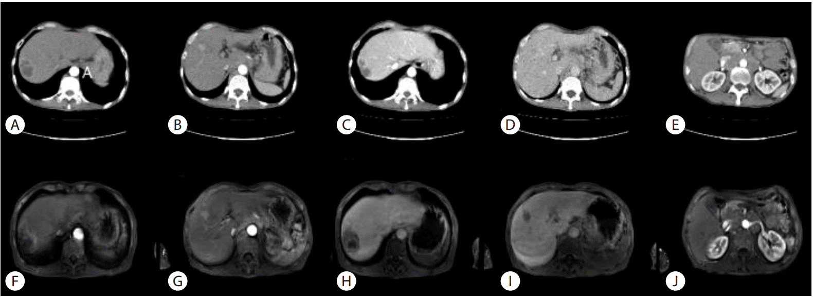
Abdominal computed tomography (CT) and magnetic resonance (MR) imaging. CT images shows several masses with or without central necrosis at the liver S6, S7 and 8 (diameter of the largest one: 5 cm). The masses shows early enhancement (A, B) and delayed wash out (C, D). Multiple enlarged lymph nodes (LNs) are noted along the celiac axis, common hepatic artery, portocaval, paraaortic, aortocaval, and retrocaval areas (E). MR images also shows several masses. The masses shows early enhancement (F, G) and delayed wash out (H, I). Multiple enlarged LNs are also noted along the celiac axis, common hepatic artery, portocaval, paraaortic, aortocaval, and retrocaval areas (J).

Positron emission tomography (PET)-CT and chest CT. (A) PET-CT shows mildly increased fluodeoxyglucose uptake in the liver dome (SUVmax : 3.6). (B, C) Multiple FDG uptake is noted in retroperitoneal, gastrohepatic, and left supraclavicular lymph nodes. (D) Cavitary pulmonary nodules is also noted in both upper lobe. (E, F) Chest-CT shows multiple thick-walled cavitary lesion, branching linear opacities and clustered centrilobular nodules at both upper lobe.
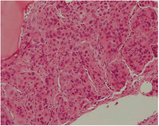
Bone marrow biopsy histology (H&E stain, ×200). Tumor cells are infiltrating bone marrow space. They have polygonal ample cytoplasm with round nuclei showing conspicuous nucleoli and grow in trabecular pattern. Morphologically, it is well consistent with hepatocellular carcinoma.
DISCUSSION
The bone is one of the common sites for metastasis in HCC, although bone marrow metastasis from HCC is rarely reported and its frequency is not well known. Bone marrow metastasis from solid organ malignancies generally occurs at the advanced stage. It worsens the quality of life, causes compression fracture, cancer pain, disseminated intravascular coagulation (DIC), and death [14,15]. The above advance stage is sometimes called disseminated carcinomatosis of the bone marrow (DCBM). DCBM is an infiltrative metastasis in the bone marrow and is characterized by anemia, back pain, and bleeding tendency. In addition, the disease is often associated with DIC and both conditions are associated with poor prognosis. Although, at the time of diagnosis, cytopenia was a frequent finding, especially anemia and thrombocytopenia [12], there is only one case that report DCBM associated with HCC [13].
Almost all types of malignant tumors can metastasize to the bone marrow [16-18]. Lung, breast, and prostate malignancies in adults, and neuroblastoma and rhabdomyosarcoma in children are the most common nonhematological malignancies frequently metastasize to the bone marrow [16]. The detection depends on the stage of the malignancy and the other sites of the metastases. Therefore, bone marrow metastasis affects the stage of the malignancy and overall survival. Bone marrow biopsy is needed to confirm the diagnosis. The confirmation of diagnosis does not always affect the treatment or prognosis of disease, because clinical course is rapid and it is usually accompanied by DIC. However, it is also reported that the early diagnosis and chemotherapy improved DIC and improved the survival for a few months [19]. Although bone marrow metastasis is accompanied by bone metastasis in the majority of cases [16,17], in our case, there was no evidence of bone metastasis on abdomen CT and PET-CT, which let us not suspect the bone marrow metastasis at the time of HCC diagnosis.
In conclusion, although HCC accompanied by underlying cirrhosis usually showed anemia, thrombocytopenia or pancytopenia and diagnosis of bone marrow metastasis may be rarely meaningful to treatment and prognosis in the rapidly progressive case, physicians should always suspect the bone marrow involvement in cancer patients with anemia and thrombocytopenia. Therefore, a bone marrow examination should be performed without delay to diagnose and treat at an earlier phases of the disease.
Acknowledgements
This work was supported by the National Research Foundation of Korea (NRF) grant funded by the Korea government (MSIP) (NRF-2015R1A5A2009656).
Notes
Conflicts of Interest: The authors have no conflicts to disclose.
Abbreviations
CT
computed tomography
DCBM
disseminated carcinomatosis of the bone marrow
DIC
disseminated intravascular coagulation
Hb
hemoglobin
HCC
hepatocellular carcinoma
LN
lymph node
MRI
magnetic resonance imaging
PET
positron emission tomography