


INTRODUCTION
Surveillance screening for hepatocellular carcinoma (HCC) has been accepted as standard care for patients with chronic liver disease who are at risk of developing HCC [1-5]. Ultrasonography (US) has become an established primary surveillance tool for the detection of HCC, given its non-invasiveness, widespread availability, acceptance by patients and physicians, and relatively low cost.
The US features of HCC, other hepatic malignancies such as metastasis or cholangiocarcinoma (CC), and benign lesions such as hemangioma have been sporadically described in literature [6-14]. However, there is a lack of uniformity in descriptive terminology for US features, which can limit its application. Therefore, the creation of a lexicon is advocated for better communication of radiological features, in order to establish standard terminology for use in daily practice and clinical research. In addition, to the best of our knowledge, no previous study has investigated the possibility of using US features for differentiating between benign and malignant lesions in a clinical setting of surveillance US for HCC.
The purposes of our study were to propose a lexicon for liver US and identify radiological features indicative of benign or malignant lesions during surveillance US.
MATERIAL AND METHODS
Patients
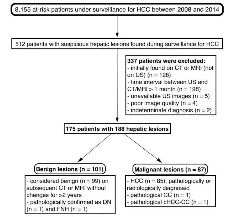
This study was approved by our institutional review board, and informed patient consent was not required. Between January 2008 and December 2014, 8,155 patients at high risk for HCC underwent surveillance US more than once at an academic tertiary referral hospital in Seoul, Korea. Liver US and serum alpha fetoprotein (AFP) assay are routinely used in conjunction for HCC surveillance at our institution. Computed tomography (CT) or magnetic resonance imaging (MRI) is occasionally performed for the purpose of surveillance at the discretion of the clinician. Upon reviewing the medical records and imaging data of the 8,155 patients, 512 patients who were suspected for HCC during surveillance were identified. Of the 512 patients, 337 were excluded for the following reasons: suspected HCC was initially identified upon CT or MRI instead of US (n=128); the time interval between surveillance US and subsequent CT/MRI was longer than 1 month (n=198); images could not be retrieved (n=5); US image quality was too poor to allow evaluation (n=4); and hepatic lesions remained indeterminate (n=2). The final study cohort consisted of 175 patients with 188 nodules (benign, 101; malignant, 87).
Of the 101 benign lesions, while 2 were pathologically confirmed to be dysplastic nodules and focal nodular hyperplasia by biopsy (n=1, each), the remaining 99 were either not visualized (n=54) upon subsequent imaging studies or considered benign (n=45) based on the absence of changes in subsequent dynamic contrast-enhanced CT or MR images acquired during follow-up evaluations for over 2 years. Of the 87 malignant lesions, 85 were determined to be HCCs based on pathological findings or visualization of hallmark radiological findings (arterial enhancement and venous washout) upon subsequent dynamic imaging studies, while 2 were determined to be other hepatic malignancies (pathologically diagnosed CC and combined HCC-CC). Schematic representation of patient selection and diagnostic results are presented in Figure 1.
Clinical and laboratory data and pathology reports of these patients, including patient demographics, etiology of chronic liver disease, serum AFP levels, and pathological findings, were retrospectively reviewed. The reference value for serum AFP concentration used at our institution is <9 ng/mL.
Ultrasonography and image analysis
Abdominal US for HCC surveillance was performed using commercially available ultrasound machines (Pro-Sound Alpha10 or Pro-Sound F75, Hitachi Aloka Medical, Tokyo, Japan; ACUSON S2000, Siemens Medical Solutions, Mountain View, CA, USA; iU22, Philips Medical Systems, Best, The Netherlands) with 5-MHz curved-array transducers. Image acquisition was performed according to our established protocol. Patients with suspected portal vein thrombosis underwent grayscale as well as Doppler imaging.
All US images were retrieved from a Picture Archiving and Communication System (Centricity, Version 2.0, GE Healthcare, Barrington, IL, USA). Two abdominal radiologists (M.S.P. and C.A., with 19 and 6 years of experience in acquisition and interpretation of abdominal US images, respectively) reviewed the literature on the US features of hepatic lesions and recorded relevant lexicons to subsequently create our own lexicon, which was applied for the evaluation of the cases included in the present study.
Based on the newly defined lexicon, two other abdominal radiologists (J.Y.L. and N.S., with 6 and 7 years of experience in abdominal US) performed blinded and independent reviews of the US images included in this study. They also evaluated the background liver parenchyma to classify it as cirrhotic or non-cirrhotic. Prior to the independent review process, they underwent training for the use of the lexicon, during which they reviewed 20 cases in consensus; these cases were not included for further analysis in our study. All US images meant for independent review were deidentified in a random order by one investigator (C.A.) and transferred to a separate workstation (Intellispace Portal 5.0, Philips, Best, The Netherlands) for blinded evaluation. Data regarding the size and number of hepatic lesions were retrieved from prospective US reports without reevaluation. Following the first independent image analysis, the interobserver agreement was evaluated, and the two reviewers drew conclusions regarding discordant results by consensus.
Statistical analysis
Comparison of variables between patients with benign and malignant hepatic lesions was performed using the Mann-Whitney U test for continuous variables and the chi-square or FisherŌĆÖs exact test for categorical variables. Correlation between US features and benignity/malignancy was determined using the chi-square or FisherŌĆÖs exact test.
The associations between US features and malignancy were determined by univariate and multivariate logistic regression analyses, and odds ratios (ORs) with 95% confidence intervals (CIs) were calculated for each of the features. Variables with alpha values <0.1 in univariate analysis were further evaluated by multivariate logistic regression analysis, where, ORs for tumor size and AFP were calculated per increments of 1 mm and 10 ng/mL, respectively.
Interobserver agreement was expressed by CohenŌĆÖs kappa or weighted-kappa coefficient (╬║). A kappa statistic value of 0.8-1.0 was considered to indicate excellent agreement; 0.6-0.79, good agreement; 0.40-0.59, moderate agreement; 0.2-0.39, fair agreement; and 0-0.19, poor agreement.15 Two-sided P-values <0.05 were considered statistically significant. All statistical analyses were performed using the SAS 9.2 software (SAS Institute Inc., Cary, NC, USA).
RESULTS
Baseline patient characteristics
The demographic characteristics of the 175 patients (male, 119; female, 56; median age, 57 years; range, 27-84 years) included in this study are shown in Table 1. While 81 patients were diagnosed as having HCC or other malignancies, the remaining 94 had only benign lesions. Patients with malignant hepatic lesions were older (median age, 57 years vs. 54 years; P<0.001), more likely to be carriers of hepatitis B virus (HBV; 82.7% vs. 59.6%; P<0.001), and exhibited greater maximum lesion diameters (median diameter, 3 cm vs. 1.8 cm; P<0.001) and higher serum AFP levels (median AFP level, 10.27 ng/mL vs. 3.19 ng/mL; P<0.001) than those with benign lesions. There were no significant differences between the two patient groups in terms of sex (P =0.255), background liver (P =0.302), or number of suspicious lesions identified on surveillance US (P =0.78).
Lexicon for ultrasonographic evaluation of hepatic lesions
The schematic drawing and description of our lexicon for liver US are presented in Figure 2.
The lexicon has four categories:
1) Morphology ŌĆö nodular with indistinct margin, simple nodular, multinodular confluent, or infiltrative
2) Rim ŌĆö none, hyperechoic, thin (<2 mm) hypoechoic, or thick (Ōēź2 mm) hypoechoic (Figs. 3 and 4)
3) Echogenicity ŌĆö homogeneously hyperechoic, homogeneously isoechoic, homogeneously hypoechoic, heterogeneous, or mosaic appearance
4) Posterior acoustic enhancement ŌĆö absent, present, or non-assessable (in case of lesions located in the posterior subcapsular portions of the liver)
Ultrasonographic features of benign and malignant hepatic lesions
The results of image analysis are presented in Table 2. Benign hepatic lesions were more likely to exhibit no rim (P<0.001) and homogeneous hyperechogenicity (P<0.001) than malignant lesions, while the latter were more likely to exhibit multinodular confluent morphology (P =0.02), thick hypoechoic rim (P<0.001), heterogeneous echogenicity (P <0.001), mosaic appearance (P =0.04), and posterior acoustic enhancement (P<0.001) than benign lesions. Interobserver agreement for morphology (╬║=0.36) was fair, while those for rim (╬║=0.427), echogenicity (╬║=0.549), and posterior acoustic enhancement (╬║=0.543) were moderate.
The results of univariate and multivariate logistic regression analyses (Table 3) revealed larger tumor size (OR, 1.12; 95% CI, 1.06-1.183; P<0.001), multinodular confluent morphology (OR, 7.712; 95% CI, 1.053-56.465; P =0.044), thick hypoechoic rim (OR, 5.878; 95% CI, 2.681-12.888; P<0.001), and posterior acoustic enhancement (OR, 3.077; 95% CI, 1.237-7.655; P =0.016) to be independent factors associated with malignant hepatic lesions. None of the US features were significantly associated with benign lesions.
Subgroup analysis according to tumor size
Prevalence of malignancy according to tumor size is presented in Table 4. Of the 188 evaluated lesions, 14 (7.4%) were subcentimeter (<1 cm) lesions, 62 (33%) were 1-2 cm in size, 57 (30.3%) were 2-3 cm, and 55 (29.3%) were 3 cm or larger. None (0%) of the subcentimeter lesions, 14 (22.6%) of the 1-2 cm lesions, 30 (52.6%) of the 2-3 cm lesions, and 43 (78.2%) of the lesions Ōēź3 cm were malignant.
The results of subgroup analysis of lesions <2 cm revealed that none of the US features were significantly associated with malignancy or benignity (Table 5). Furthermore, US features favoring malignancy were rarely observed in small lesions; among the 14 small (<2 cm) malignant lesions, thick hypoechoic rim, heterogeneous echogenicity, mosaic appearance, and posterior acoustic enhancement were observed in none or only a couple of cases (Table 5). Logistic regression analysis could not be performed because the frequencies of potentially significant US features were too low.
DISCUSSION
In the present study, size and three morphological features including multinodular confluent morphology, thick hypoechoic rim, and posterior acoustic enhancement were found to be significantly associated with malignancy. Multinodular confluent morphology, thick hypoechoic rim, and posterior acoustic enhancement were reported as morphological features suggestive of malignancy over two decades ago [8,10,12,14]. In spite of the recent technological developments in US, characteristic features suggestive of malignancy have remained unchanged. However, in our study, these features were mostly observed in large lesions. In addition, in case of hepatic lesions <2 cm in size, none of the US features exhibited significant association with benignity or malignancy.
All the international guidelines clearly state that US is a surveillance tool, not diagnostic [1-5]. According to the current guidelines, short-term follow-up is recommended for a hepatic lesion smaller than 1 cm found on surveillance US, while for a hepatic lesion larger than 1 cm, dynamic contrast-enhanced CT or MRI is recommended as a recall policy irrespective of US features. Our results support the recommendation by the current guidelines; in our study, all of the subcentimeter nodules were benign, and the potential for malignancy increases by more than 20% with the increase in the size of lesions beyond 1 cm, with any US feature unable to differentiate between small HCC and benign lesion.
Previous studies reporting that hypoechoic rim is suggestive of malignancy have not taken the thickness of the rim into account [9,10,12]. To reflect the evolution of technology, we divided the hypoechoic rim category into two subcategories ŌĆö thin and thick. In our study, thick hypoechoic rim was significantly associated with malignancy, while thin hypoechoic rim exhibited no significant association. Thin hypoechoic rims observed around benign lesions are more likely to be pseudo-rims, i.e., Mach bands rather than true rims, created because of an optical effect at margins between areas of different echogenicities [16]. In contrast, hyperechoic rim with partially hypoechoic internal pattern has been reported as being specific for hepatic hemangioma [11,13]. In our study, 5 of 6 lesions with hyperechoic rims were benign, which suggests that hyperechoic rim might be indicative of benign lesions; however, the statistical significance of this association could not be established in our study, possibly because of the low frequency of occurrence of hyperechoic rims.
A major limitation of this study is that we retrospectively reviewed US images acquired by a heterogeneous group of US operators, including inexperienced ones. Therefore, the results of our study might not be relevant when prospectively applied in different settings. Another limitation could be that the difference of size distribution between benign and malignant lesions, which could affect the results. Our results showed that none of the US features was found to be significantly associated with benignity or malignancy in case of small (<2 cm) hepatic lesions. Among the 76 small nodules, only 14 (18.5%) nodules were malignant.
In conclusion, for hepatic lesions larger than 2 cm, some US features might be suggestive of malignancy. We proposed a lexicon that may be useful for surveillance US.


 PDF Links
 PubReader
 ePub Link
 Full text via DOI
 Full text via PMC
 Download Citation
 Print
Print


